LS-2-P-3151 Piecemeal degranulation is the main secretory process of eosinophils during the experimental acute Schistosoma mansoni infection
Schistosomiasis is a chronic disease caused by trematoda Schistosoma mansoni that triggers granuloma formation [1]. Eosinophils, cells of the innate immune system, are recruited and migrate to sites of granulomatous response playing effectors functions against parasite eggs through the release of secretory granule-derived proteins [2]. These granules exhibit an ultrastructurally unique morphology, with a crystalline core, and store a significant number of cytokines and cationic proteins in preformed pools within them [3]. Since little is known about the processes of secretion of these cells in response to helminthic infections, this work aimed to investigate the secretory processes involved in the eosinophil secretion during the experimental infection with Schistosoma mansoni. Female Swiss mice (n=6) were infected with 100 cercariae of S. mansoni percutaneously. Animals were euthanized (animal ethical approval CEUA/FIOCRUZ # LW-32/2012) after 55 days post-infection (acute phase) and liver fragments were processed for light microscopy (LM), conventional transmission electron microscopy (TEM), and immunogold electron microscopy using a pre-embedding approach for detection of Major Basic Protein (MBP), the major cationic protein stored within eosinophil secretory granules. LM revealed the presence of high number of infiltrating eosinophils surrounding the S. mansoni eggs into hepatic granulomas of infected mice, some of them in close contact with the S. mansoni eggs surface (Fig. 1). TEM revealed distinct eosinophil degranulation processes in the S. mansoni-infected livers, such as classical granule exocytosis and, mainly piecemeal degranulation (PMD). PMD (Fig. 2) was characterized by morphological changes of specific granules (enlargement, reduced electron-density, core disarrangement and coarse granule matrix) and presence of a high number of cytoplasmic vesicles, indicative of a vesicle-mediated transport of granule-stored products. Granules undergoing PMD were immunolabeled for MBP (Fig. 3). This means that the secretion of eosinophil products during the acute infection is occurring through mobilization and release of specific molecules. Altogether, our findings confirm that eosinophils are key cells in the S. mansoni acute infection and identify PMD as a major process of secretion in response to the infection. Moreover, the understanding of all events and mechanisms governing differential sorting, packing and secretion of granule-stored mediators may be also fundamental to the goal of specifically blocking eosinophil secretion as a therapeutic strategy.
References:
[1] Gryssels et al. (2012). Infect Dis Clin N Am, 26: 383–397.
[2] Shamri et al. (2011). Cell Tissue Res, 343: 57-83.
[3] Melo & Weller (2010). Histol Histopathol, 25: 1341-1354.
Supported by CAPES, CNPq and FAPEMIG.
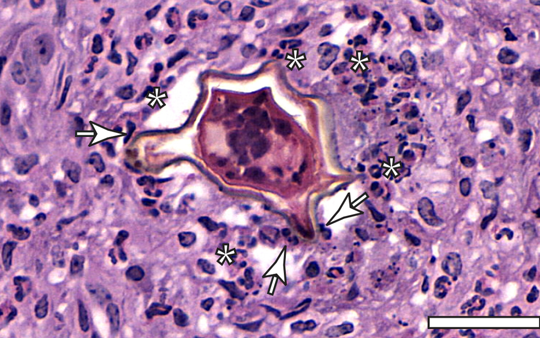
Fig. 1: A hepatic granuloma shows infiltrating eosinophils (*) surrounding and in close contact with the S. mansoni egg (arrows). Livers from S. mansoni-infected mice were processed for light microscopy and 5μm-thickness sections were stained with hematoxylin-eosin (HE). Bar: 30µm. |

Fig. 2: Electron micrograph of an eosinophil in process of piecemeal degranulation (PMD). Secretory granules show morphological changes (*) – enlargement and core disarrangement - indicative of a vesicle-mediated transport of granule-stored products. Vesicles (arrows). Livers from S. mansoni-infected mice were processed for TEM. N: nucleus. Bar: 1.3μm. |
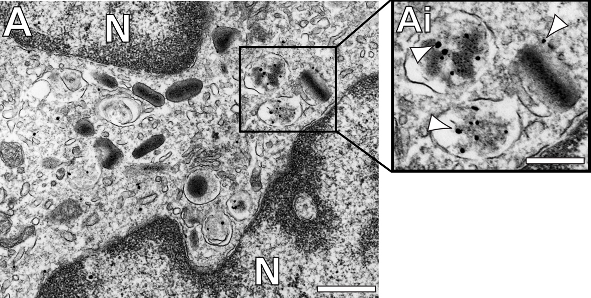
Fig. 3: (A, Ai) Eosinophil secretory granules undergoing PMD are immunolabeled for Major Basic Protein - MBP (arrowheads) (A). Liver fragments were immunolabeledd for MBP, using pre-embedding immunogold electron microscopy. N: nucleus. Bar: (A) 1.3μm; (Ai) 0.65μm. |
|