IT-16-P-2955 Removing the effects of elastic and thermal scattering from spectrum images in scanning transmission electron microscopy
Scanning transmission electron microscopy (STEM) has proven to be a powerful tool for the acquisition of atomic-column-resolved elemental maps using electron energy-loss spectroscopy (EELS) and, more recently, energy-dispersive x-ray (EDX) spectroscopy. Furthermore, in EELS, given the ability to study how spectra change on the atomic scale, there has also been much interest in mapping not only the positions of atoms, but also their bonding states by studying changes in the fine structure of EELS data [1-3]. Specifically, atomic-resolution oxygen EELS data in transition metal oxides can potentially provide information about the entire electronic structure of a material since oxygen atoms bonded to different transition metals will display distinct spectra, reflecting the local bonding state. In atomic-resolution EDX, given the localized nature of the interaction potential involved and that such maps are not further complicated by subsequent elastic and thermal scattering after ionization, there is huge promise in quantitatively measuring elemental densities at atomic resolution.
However, due to the complex elastic and inelastic scattering of the electron probe, direct qualitative and quantitative interpretation of both elemental and bond maps is difficult. In bond mapping, the scattering of the electron probe causes the spectra from inequivalently bonded atoms to become mixed, and the features that distinguish them to become ambiguous. In elemental mapping, the highly non-linear response to atomic-column densities due to electron scattering denies any direct correspondence between signal and atomic-column densities [4].
Recently, a method has been developed to remove the effects of elastic and thermal scattering from spectrum images [5]. Using the quantum excitation of phonons model [6], the cross-section expression for inelastic scattering in STEM may be expressed as an inverse problem, and the elastic and thermal scattering deconvolved from experimental data. Here we show applications of this method in both EELS [7] and EDX [8] data of SrTiO3.
[1] DA Muller et al, Science 319 (2008) 1073
[2] H Tan et al, PRL 107 (2011) 107602
[3] M Haruta et al, APL 100 (2012) 163107
[4] BD Forbes et al, PRB 86 (2012) 024108
[5] NR Lugg et al, APL 101 (2012) 183112
[6] BD Forbes et al, PRB 82 (2010) 104103
[7] MJ Neish et al, PRB 88 (2013) 115120
[8] G Kothleitner et al, PRL 112 (2014) 085501
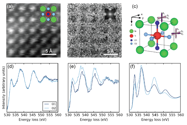
Fig. 1: (a) HAADF map of SrTiO3 [001] (projected structure inset). (b) EELS map using the O K edge (potential obtained from inversion inset). (c) SrTiO3 structure showing the two inequivalent O atoms in the [001] projection. Spectra from inequivalent O columns obtained from (d) background-subtracted data, (e) after inversion and (f) Wien2k calculation. |
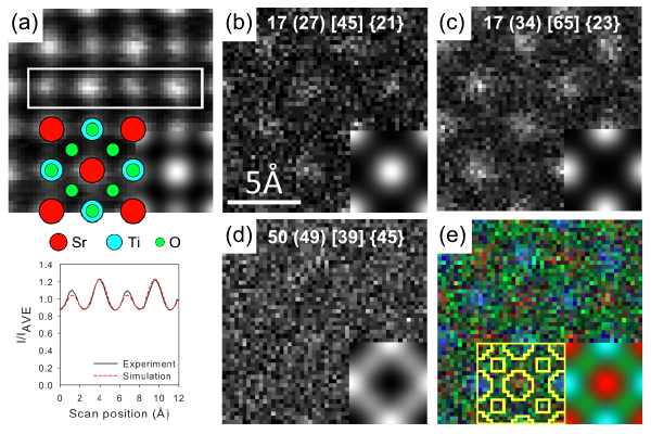
Fig. 2: SrTiO3 [001] experimental and simulated (inset) (a) HAADF (structure overlayed) and EDX (b) Sr K, (c) Ti K (d) O K edge maps. Numbers inset show atomic densities (atom/nm3) obtained from: the ideal structure (averaging the entire EDX map) [averaging specific columns] {inversion}. (e) EDX colour composite with masks used for column specific average. |