LS-3-P-2479 Living cells toward electron microscopy? Let’s do CLEM, it can be easy!
Correlative light and electron microscopy (CLEM) aims at combining the large field of view and chemical specificity of fluorescence microscopy with the high resolution ultra-structural details revealed by electron microscopy. CLEM can be extremely powerful in extending electron microscopy analysis to rare events that are impossible to target based on EM morphology alone. Furthermore, efficient workflow solution can speed up complicated experiments by extended automation of different steps along the workflow.
Here, we present different ways of using fluorescence to efficiently target areas for 3D analysis using AutoSlice&View. In the first experiment we started from an already resin embedded tissue and imaged with both modalities. This was possible thanks to a protocol that has both the fluorescence signal and contrasting agent in the embedded block. Due to purely image-based correlation, it was straightforward to directly target a specific area identified by the fluorescence signal without the use of fiducial markers of any kind. The second experiment covers imaging from living cells in the light microscope, to resin embedded cells in the electron microscope. Here we could identify a living cell with an endocytic event occurring through light microscopy and follow that very structure all the way through the sample preparation until the final high resolution 3D electron microscopy. In this second workflow we also introduced automation of the sample preparation steps including measures for efficient relocation procedures.
These experiments clearly highlight the strong potential of using correlative approaches to target small sub-volumes in larger volumes for efficient AutoSlice&View acquisition of 3D electron microscopy data. The use of a flexible software framework is needed to accommodate different workflows, while automation of sample preparation steps will help to cut down the complete experiment time tremendously.
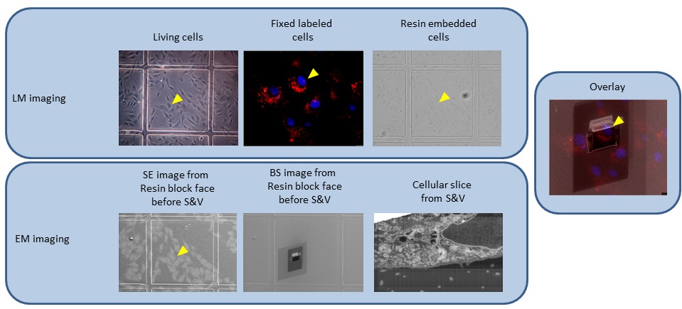
Fig. 1: Images through the complete experiment: from living cells in the light microscope, to fixed embedded cells first in the light microscope and then in the FIB/SEM. The yellow arrowhead indicates the cell of interest, which has been tracked throughout the workflow |

Fig. 2: Images through the complete experiment. From top to bottom images collected in the light microscope first and in the FIB/SEM then. In the middle panel are displayed the overlays between the LM and EM images used to correlate. The yellow arrowhead point at the cell followed throughout the workflow |