IT-4-P-2405 Application of Multi-Tilt Specimen Stage for Advanced Electron Channeling Contrast Technique
New developments for a selected area electron channeling pattern (SACP) acquisition and electron channeling contrast imaging (ECCI) in the scanning electron microscope (SEM) are presented. Novel approaches for electron channeling contrast formation are introduced using multi-axial multi-tilt specimen stage.
The multi axial piezo-driven specimen stage allows high precision movement with all six degrees of freedom. Among other applications, it allows bi-axial tilting around any selected point on the sample. Such a technology enables precise selection of beam to surface angle which is the core principle of electron channeling contrast formation, as shown in Fig. 1.
The well-known "rocking beam" technique for electron channeling pattern (ECP) described e.g. in [1] is based on a special mode of scanning in the SEM. Limitation of this technique is given by spherical aberration of the objective lens, which restricts its use mainly to single crystals. A dedicated Cs corrected rocking beam mode was developed for TESCAN field emission microscopes for acquisition of SACP from a very small area as shown in Fig. 2. The practical use of this correction was demonstrated on polycrystalline samples in [2]. Further extension of the rocking angle by the use of stage tilt was also tested.
Specific properties of the ECCI technique in SEM for the observation of near surface defects are explained. The relation of ECP to the formation of ECCI is crucial for understanding the whole ECCI phenomenon. The oriented ECCI technique for reaching suitable diffraction condition as described in [3] was applied. Advantage of combination of Cs corrected SACP for oriented ECCI technique is shown. Newly, the use of precise bi-axial specimen tilt for oriented ECCI is demonstrated in Fig. 3.
Furthermore, new techniques for ECCI contrast improvement, such as color coded multi-axial specimen tilt, are introduced. The sample tilt angle is coded according to HSV color or RGB model to improve the informational depth of the micrograph (see Fig. 4).
References:
[1] A J Wilkinson et al, Micron 28 No. 4 (1997) p. 279.
[2] J Dluhoš et al, METAL Conference Proceedings (2012) p.453.
[3] B A Simkin et al, Ultramicroscopy 77(1-2) (1999) p. 65.
The research has been supported by the Technological Agency of Czech Republic TE 01020233(AmiSpec)
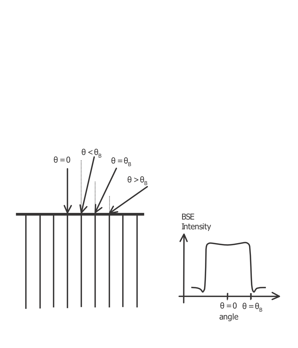
Fig. 1: Schematic diagram of forming the channeling contrast in relation to deviation from the Bragg condition. Image by Wilkinson et al. [1]. |
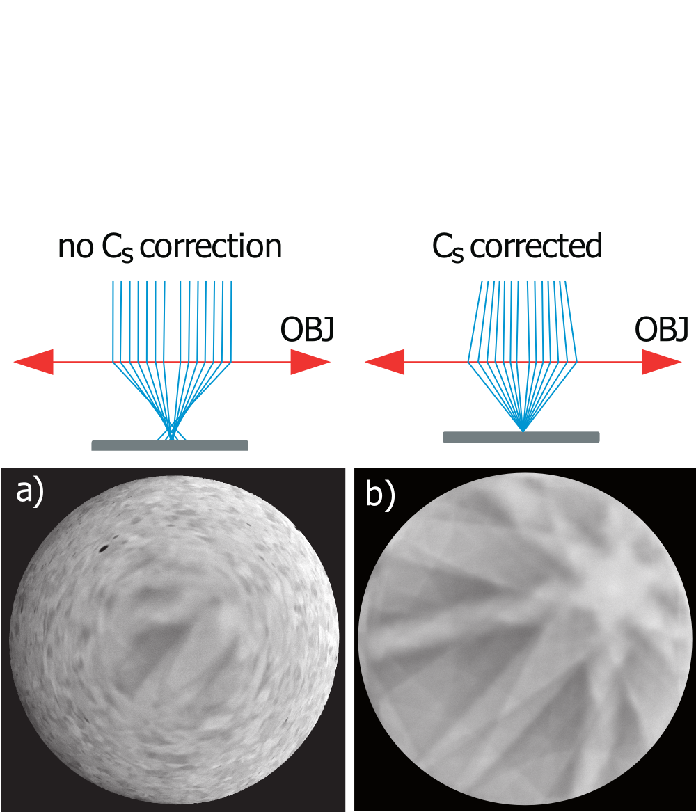
Fig. 2: Comparison of ordinary rocking beam mode without correction of spherical aberration (left) and a Cs corrected ECP mode (right). Images taken on polycrystalline stainless steel with grain size about 20 µm. |
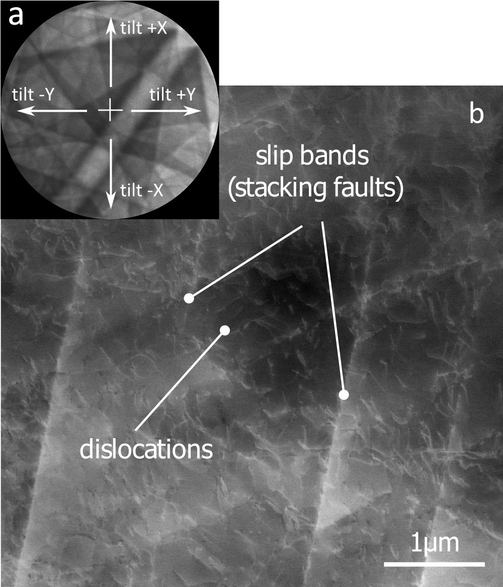
Fig. 3: ECCI imaging with the use of SACP a) navigation to diffraction condition on SACP (edge of the band, using a multi axial stage tilt. b) ECCI image of crystal defects |
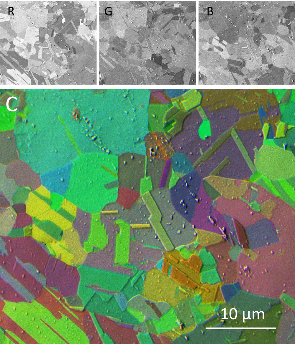
Fig. 4: Composite ECCI micrograph of copper sample with randomly oriented grains using the color coded tilt of the stage. R,G,B – images acquired with specimen tilt from -5° to +5°. |