MS-4-O-1559 Structure-Property Relationships in Fe-Mn Austenitic TRIP/TWIP Steels Determined With Conventional and Aberration-Corrected Transmission Electron Microscopy
A new class of austenitic steels stabilized with high Mn contents (instead of Ni) exhibits exceptional mechanical properties, such as large energy absorption and high work-hardening rate, owing to secondary deformation mechanisms such as mechanical twinning-induced plasticity (TWIP) and martensitic transformation-induced plasticity (TRIP) favored for low stacking-fault energy (SFE) [1]. The interaction of dislocations with twin boundaries and martensite interfaces during mechanical deformation enhances the work hardening, i.e., a dynamic Hall-Petch effect, with total elongations exceeding 70% and ultimate tensile strengths in the GPa regime.
In this investigation, the SFE and deformation mechanisms of Fe-(22,25,28)Mn-3Al-3Si (wt%) austenitic steels have been studied with a combination of conventional and advanced electron microscopy to make correlations with the work-hardening behavior and mechanical properties. The SFE measurements employed weak-beam dark-field (WBDF) imaging to measure the separation of partial dislocations. Figure 1 is a WBDF image from Fe-22Mn-3Al-3Si recorded with a Philips CM20. Using single-crystal elastic constants to determine the effective shear modulus on the (111) slip plane and effective Poisson’s ratio, the SFE energies for the 22, 25 and 28% Mn alloys are 15 ± 3, 21 ± 3 and 39 ± 5 mJ/m2, respectively [2]. Deformation mechanisms were characterized by bright-field (BF) imaging of interrupted tensile tests. Figure 2 shows epsilon-martensite lath formation in the 22% Mn alloy after 10% strain. As the SFE increases, the secondary deformation changes from martensite to mechanical twining as shown in figure 3 from the 28% Mn alloy with 10% strain.
In order to better understand the role of twin boundaries and martensite interfaces on work hardening, high-resolution imaging (HRTEM) using an image-corrected FEI-Titan provides both qualitative and quantitative information about the strain fields at these interfaces. Figure 4 is an HRTEM image from the 28% Mn alloy of a twin boundary in a sample with 20% strain. The twin plane exhibits a lack of mirror symmetry which could contribute to the strong work-hardening effect. Quantification of the strain fields at these interfaces is currently ongoing using real-space strain measurements. The relatively thick electropolished samples (t/λ maps indicate that t~ 20 nm) and 20% deformation limit image quality. Improved images may be obtained with planned aberration-corrected STEM imaging.
References
[1] O. Grassel, L. Kruger, G. Frommeyer, and L. W. Meyer, Int. J. Plasticity,16(2000) p.1391
[2] D.T. Pierce, et al., Acta Mater 68 (2014) 238-253
Financial support from the NSF DMR 0805295 and the SFB 761 “Steel –ab initio” and research at the Ernst Ruska Centre are gratefully acknowledged.
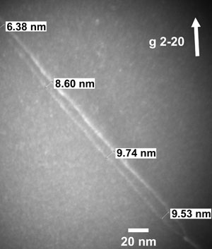
Fig. 1: Weak Beam Dark Field image of partial dislocations in Fe 22Mn-3Al-3Si for stacking fault energy (SFE) measurements (sg = 0.15 nm-1). |
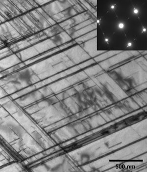
Fig. 2: Bright Field of the Fe 22Mn-3Al-3Si alloy after 10% deformation exhibiting two variants of epsilon martensite formation. |
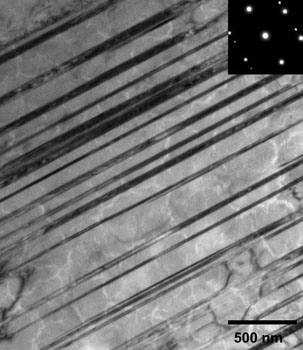
Fig. 3: Bright Field image of the Fe 28Mn-3Al-3Si alloy after 10% strain revealing multiple deformation twins. |
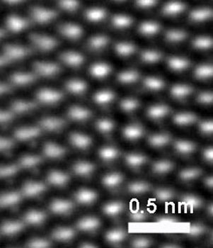
Fig. 4: High Resolution TEM image of a twin boundary in the Fe 28Mn-3Al-3Si alloy after 20% deformation. |