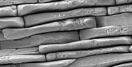
1- gel beads coated with a ruc13 coatings, magdalena parlinska, university of rzeszow, poland Weathered Rock, FEI Courtesy: Images Sperm Tails, FEI Courtesy: Images
PAN Microfibers, FEI Courtesy: Images Bacteriophage T4 Virus, FEI Courtesy: Images 2- diatom, magdalena parlinska, university of rzeszow, poland
energy filtered tem micrograph of yttria (in green) - zirconia (in red) multilayers, chanchal ghosh,  igcar, kalpakkam, india  Offretite Scagno, TESCAN 1- cvd grown diamond film, magdalena parlinska, university of rzeszow, poland
orchid root stained with acridine orange, s. r. senthilkumar, st. josephs college, india orchid root with mycorrhiza, s. r. senthilkumar, st. josephs college, india Powder metallurgy substrate, TESCAN
2- gel beads coated with a ruc13 coatings, magdalena parlinska, university of rzeszow, poland orchid root showing with idioblastic cells, s. r. senthilkumar, st. josephs college, india Calcium Sulfate Crystals, FEI Courtesy: Images
2- cvd grown diamond film, magdalena parlinska, university of rzeszow, poland Alveoli, FEI Courtesy: Images collagen fibers in cartilage, e. i. romijn, ntnu, trondheim


LABYRINTH OF MICROSCOPY

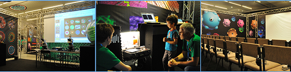
Labyrinth of Microscopy was an important IMC 2014 side-event targeted at youth and public. We introduced current microscopy and provided an insight into opportunities that microscopy provides to our life and science.

As active playing is the best way for children to absorb the complicated world of science, we installed interactive demo stages where scientists guided them through the amazing achievements of science. We demonstrated different microscopic techniques used in life sciences, medicine, nanotechnology and other fields, while the children was pass through the labyrinth and fulfiled various tasks.

poster of the Labyrinth of Microscopy (pdf)


VIDEO REPORTAGE


PHOTO GALLERY


Skinhome - samolepky na zeď Skinbook - samolepka na notebook Fabrix - pouzdra na notebooky, iPady, iPody, iPhony, mobilní telefony, hard disky, fotoaparáty