IT-10-P-6047 Multi-Axis Electron-Holographic Tomography
Electron holography has proven to be a suitable method for measuring electrostatic and magnetic fields of nanostructures in the TEM. In electron holographic tomography (EHT) [1], the intrinsic fields can be mapped in all three dimensions, thus providing quantitative access to the true inner potentials and not only their projections. However, single-axis (SA) electron tomography (ET) often suffers from loss of information in one dimension, due to the limited tilt range of common tomographic specimen holders. In Fourier space, this limitation leads to an unsampled area, the so-called “missing wedge”. Performing multi-axis ET by combining several SA tilt series of the same object, one can minimize this missing volume. In case of Dual-Axis Tomography (DA) for example (two series acquired with tilt-axes perpendicular to each other) the volume can be reduced to a “missing pyramid” [2]. Fig 1 shows the corresponding missing volumes in Fourier space, depending on the available tilt range. For usual tilt ranges of about +-70°, DA is expected to provide a much better resolution in the third dimension than SA.
Another potential benefit of Multi-Axis EHT is the possibillity to reconstruct more then only one component of the B-vector field of magnetic samples.
Here we report on the development and implementation of Multi-Axis EHT.
For the necessary alignment of the residual displacements within the tilt series, the centre of mass for the projected potential at each tilt has been determined [3]. This method has two major advantages: all tilt series are inherently aligned with respect to each other, and the (projected) tilt axis is automaticaly alligned.
For tomographic reconstruction, a self-written software program has been developed. It is based on the concept of a weighted simultaneous iterative reconstruction technique (WSIRT [4]) but is especially adapted to the peculiarities of multi-axis geometries (Fig 2). Combining all tilt series within the process of a 3D back-projection, before application of a weighting filter in 3D-Fourier space and applying the iteration loop, takes into account the projections of all tilt series at once, instead of applying the weighting and iteration loop for each 2D-slice independently with separate SA tilt series.
As an example in Fig 3 the 3D potential of barium titanate nanoparticles [5], reconstructed by DA EHT, is compared with the two corresponding 3D potentials, reconstructed from only one of the two SA tilt series of the complete dataset. In z-direction the DA tomogram exhibits sharper edges then its SA counterparts.
[1] D Wolf et al. Ultramicroscopy, 110(5) (2010), 390-399
[2] P Penczek et al. Ultramicroscopy, 60(3) (1995), 393-410
[3] S Sturm. Diploma thesis, TU Dresden (2011)
[4] D Wolf. Dissertation, TU Dresden (2010)
[5] BTO nanoparticles provided by D. Szwarcman and G. Markovich (Tel-Aviv University).
[6] Funded by the EU (ERDF) and the Free State of Saxony via the ESF project 100087859 ENano.
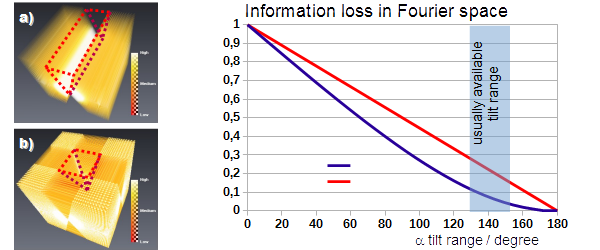
Fig. 1: Fig. 1: Missing wedge (a) and missing pyramid (b) in Fourier space and information loss according to the corresponding volumes. |
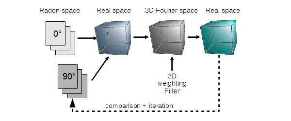
Fig. 2: Fig. 2: 3D-WSIRT algorithm reconstructing several tilt series synchronously. |
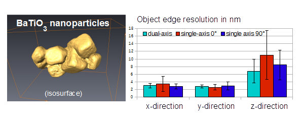
Fig. 3: Fig. 3: Dual Axis EHT reconstruction of BaTiO3 nanoparticles. The measured object edges in the tomogram are compared with single axis reconstructions of the two data subsets. |
|