IT-2-P-3462 The mini-TEM: highquality imaging and analysis of biological specimen
Traditional transmission electron microscopes are bulky and complex machines that are mostly operated by trained specialists. In this paper, we introduce the miniTEM, shown in Fig. 1, a desk-top instrument designed for imaging of biological as well as inorganic samples. The miniTEM has a high degree of automation in the microscope alignment, image acquisition, and analysis processes. The idea with the miniTEM is a small, cheap, robust, and easy to use system that requires no more training than any simple lab equipment, and can be hosted in any office or lab (even a mobile lab). Here we illustrate the imaging possibilities and show that it is good enough for quantitative and qualitative analysis.
The miniTEM microscope runs at 25 keV, which enables high-quality imaging of biological samples with a thickness up to at least 100 nm. The height of the microscope is only 70cm and it can sit and run on any desk in any lab or office space. It achieves a resolution sufficient for tasks such as virus identification in clinical samples, and morphological nanoparticle analysis. In addition to TEM functionality, the miniTEM can also run in STEM (scanning transmission electron microscopy) mode. We present here the achieved parameters of resolution, applicable sample thickness and image signal collection efficiency in both operating modes. One of the first images of inorganic nanoparticles acquired with a miniTEM microscope prototype in the TEM mode is shown in Fig. 2.
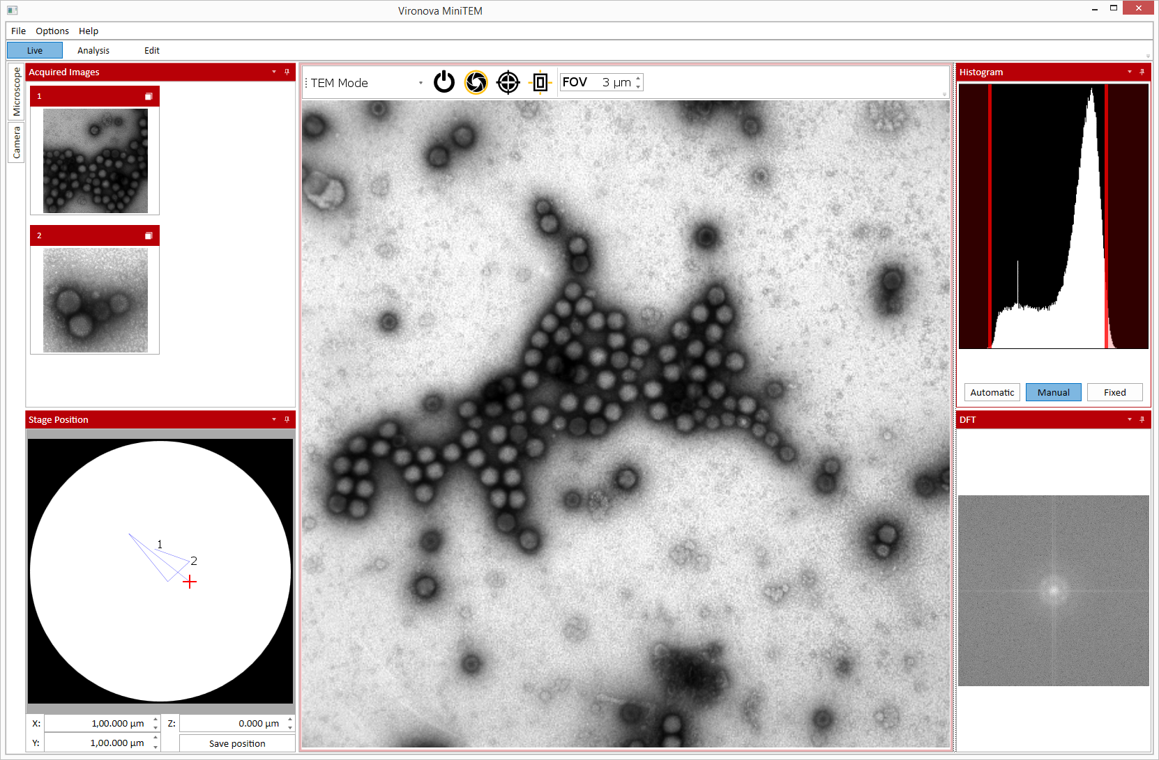
The graphical user interface is divided into three main views: live, edit, and analysis. It is developed for Windows 8, and designed for a touch screen, allowing convenient scrolling over the sample and zooming in (changing magnification). The live view, illustrated in Fig. 3, is used when manually investigating the sample by moving around, changing magnification and acquiring images. The edit view is for manually marking, drawing, measuring and annotating objects in the images. In the edit view the user can also manually correct analysis results, i.e., remove, add, and rename objects. The analysis view is where the user creates and applies automated image acquisition and/or image processing (GPU-accelerated) and analysis scripts. A graph-based interface is used to create scripts, which can be saved for future use and applied to multiple images.
This work is part of the miniTEM project funded by EU and EUREKA through the Eurostars programme.


