IT-10-P-3225 Dedicated and innovative system for tomography in the Scanning Electron Microscope
For 3D non-destructive materials characterization, two are the leading tomography techniques: X-Ray Computed Tomography and Electron Tomography implemented in TEM operated in STEM mode [1]. The first one is undoubtedly the most important for industrial applications, providing resolution of few tens of μm, for cm- to mm-scale objects, while, the second one is of great interest in many research fields, and is capable of 3D reconstruction of sub-μm-scale objects with a resolution up to 0.24 nm [2].
In this paper we will highlight the implementation and the capabilities of an alternative electron tomography system in a SEM operated in STEM mode, composed by dedicated sample holder, STEM detector and analogue/digital signal processing system.
This system aims to cover the range between the previously mentioned techniques, taking advantages of the flexibility of the SEM platform, and of the resolution and image quality capabilities of the STEM mode implemented [3]. We will show that the STEM-in-SEM tomography approach opens up the perspective for the 3D analysis of volumes up to 100 μm3, such as nanowires, carbon based nanostructures or biological specimens, with resolution up to 10 nm.
Fig. 1 shows the main building blocks of the of the innovative tomographic acquisition system. The principal constraint for the success of tomography using STEM imaging is to maintain the monotonic variation of the mass-thickness contrast over the whole tilt range. Therefore, the use of STEM for tomography requires an acquisition system capable of collecting the transmitted electron over a large and adaptable collection angles. The detector geometry with five independent circular active sectors permits to optimize the efficiencies of signal collection, as a function of the tuneable specimen-detector distance, energy and beam current. The dedicated specimen holder with rotation capability ensures the eucentricity of the observed detail and a reduced missing wedge, which are fundamental for 3D reconstruction purposes (Fig. 1a). Moreover, the dedicated signal processing system (Fig. 1b) performs faster than conventional STEM systems in the acquisition of the tilt series, preserving the amplification, conditioning and managing of the signals as required by tomographic reconstruction.
Finally, we will demonstrate the potential of the method with examples of 3D reconstruction of micro and nano-structures. In Fig. 2 is reported the tomographic reconstruction and the 3-D rendering of a bundle of human skin collagen fibers, as obtained with the first release of our dedicated system [4].
References
1. P.A. Midgley et al. J. of Ultram. 223 (2006) 185
2. M.C. Scott et al. Nature 483 (2012) 444
3. V. Morandi et al. App. Phys. Lett. 90 (2007) 163113
4. TomoSEM Project: F97I12000120007
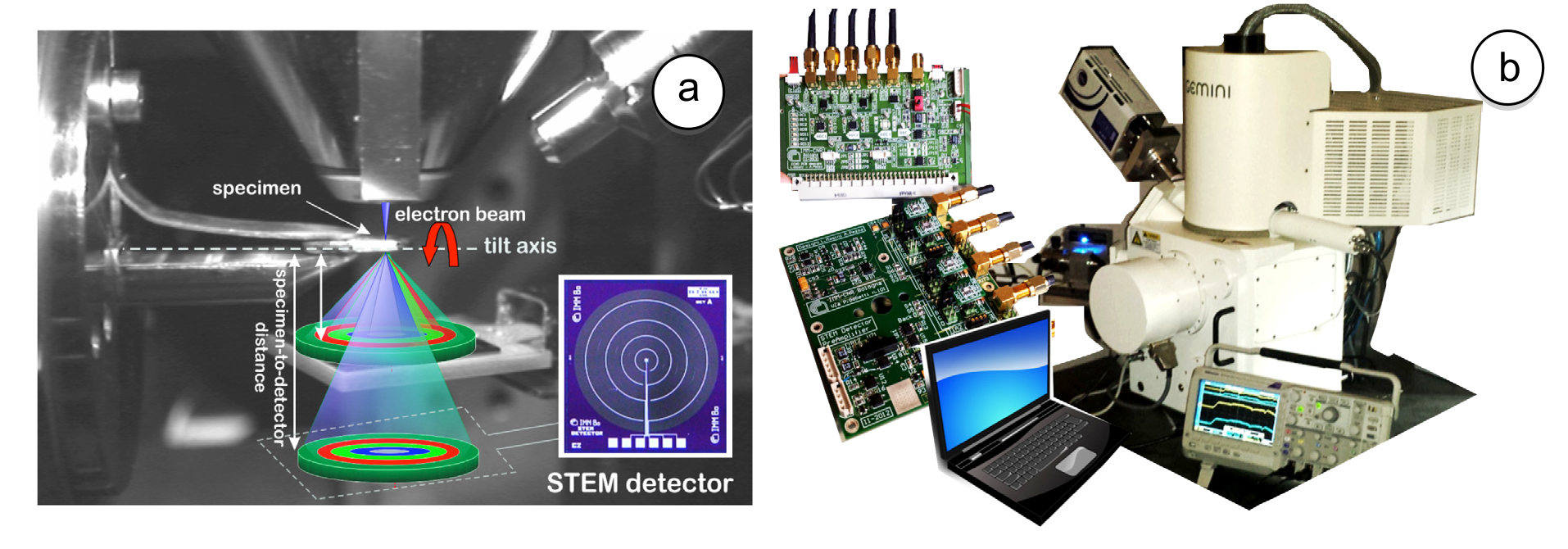
Fig. 1: (a) Setup inside the SEM-chamber: specimen-holder on motorized stage and STEM detector on a specific holder with variable working distance capability. (b) SEM platform and external mixed signal boards for amplification and managing of the signals. |
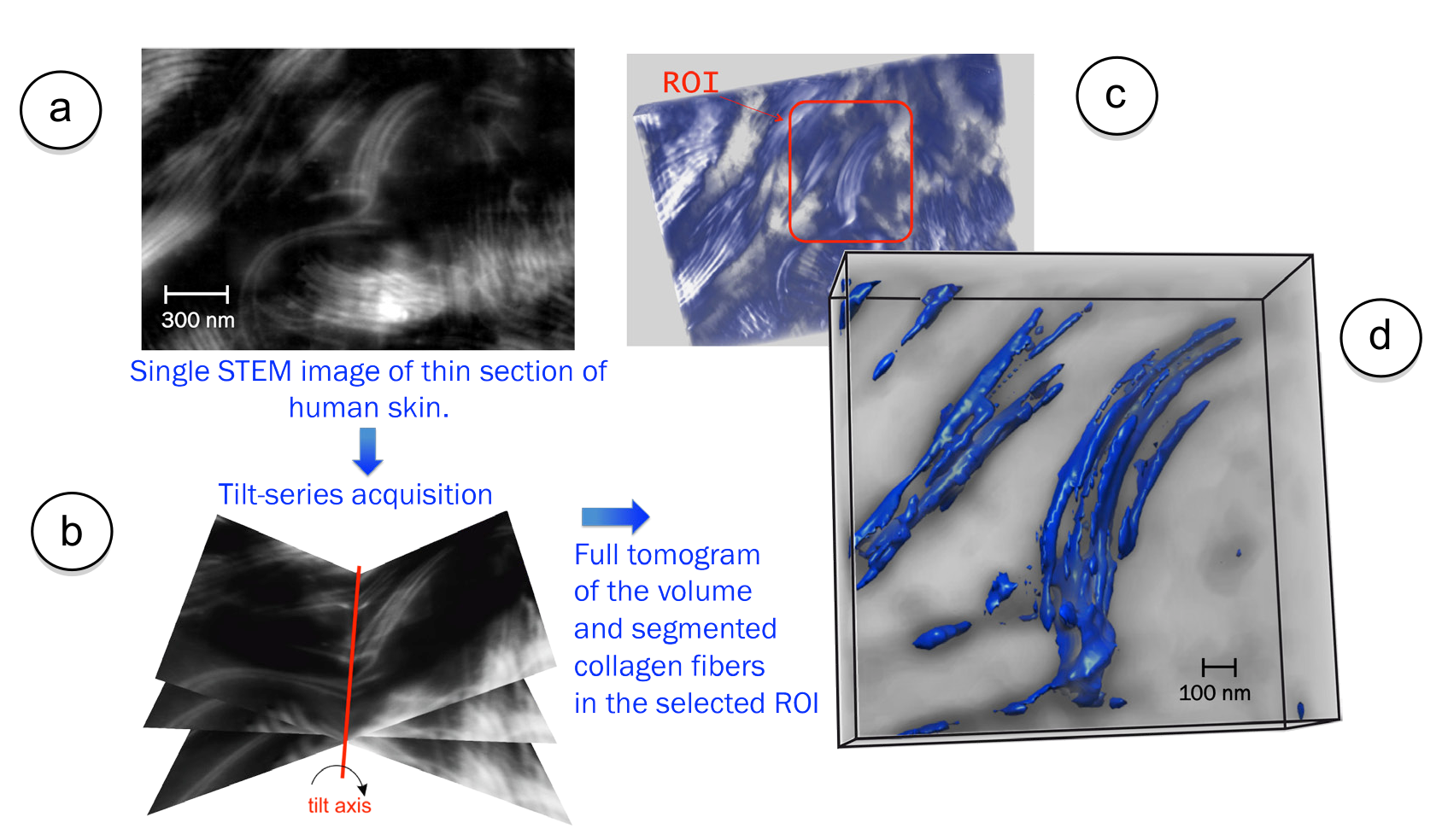
Fig. 2: Acquisition steps for a complete tomogram of a collagen fibers bundle. (a) STEM image of a human skin thin section. (b) Tilt series acquisition. (c) Reconstructed tomogram with highlighted the ROI shown in the 3-D rendering in (d). |