IT-10-P-3208 Electron tomography in the scanning electron microscope
The achievements in the implementation of electron tomography in the scanning electron microscope (SEM) and the potential of this 3-D imaging technique are summarized and discussed.
In SEM, the 3D imaging strategies consist in slice-and view assisted by FIB or microtomy, for the investigation of large specimen volumes. Differently the best resolution is pursued for relatively small volumes through TEM at high beam voltage.
The proposed implementation of electron tomography in the SEM is appropriate to the investigation at nanometric resolution of specimen volumes in the intermediate range, namely 2000 (w) x 2000 (l) x 200 (thickness) nm, as it combines the reconstruction algorithm with the signal corresponding to incoherently scattered electrons in the Scanning-Transmission (STEM) imaging mode. STEM imaging takes advantage from some peculiar characteristics of the experimental set-up [3]. This approach attains nanometric resolution and is free from aberrations caused by post-specimen imaging lenses; it also allows to collect transmitted electrons over a wide angular range [4][5]. The optimization of detector design and performance makes the contrast comply with local variations of composition or projected thickness. The bright-field component of the transmitted electrons can be effectively separated from the dark-field one, by varing the detection strategy [4].
The STEM mode preserves the monotonic variation of the signal with specimen thickness and meets the basic projection requirement for the 3-D analysis of nanowires, carbon based nanostructures or ultrastructures of biological specimens. In addition, the large value for the maximum detection angle ensures a complete detection of the scattered electrons, even in case of relatively large specimen thickness. In the case of tomography, these features are essential to maintain the proper image contrast when the specimen is rotated.
Fig. 1 shows the reconstruction of a ZnO crystalline nanostructure from a 110° tilt series at 1° step. ImageJ [6] with the TomoJ plug-in was used [7]. The disposition of the wires, their uniform section and the tapered termination are properly retrieved. Similarly, carbon-based tubes, filled with cobalt nanoclusters, were reconstructed as shown in Fig. 2. The tomogram from the STEM tilt series featuring compositional contrast, clearly shows the cobalt clusters inside the tubes.
These results demonstrate the potential of the method and optimization of the experimental set-up is under development to consolidate this technique in the set of 3-D methods of electron microscopy.
REFERENCES
1 Merli et al. Ultramic. 88 (2001) 139.
2 Morandi et al , JAP 101 (2007) 114917.
3 Morandi et al. APL 90 (2007) 163113.
4 http://rsbweb.nih.gov/ij/
5 Messaoudii et al, BMC Bioinf. 8 (2007) 288.
The authors acknowledge the finacial support from TomoSEM (F97I12000120007)
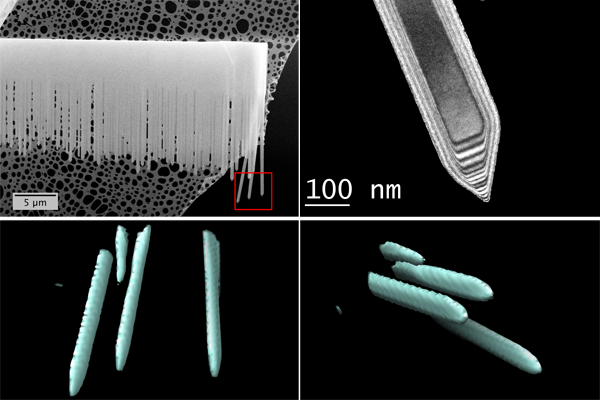
Fig. 1: Up-Left) SEM image of ZnO nanowires. The ROI is boxed. Up-Right) TEM shows the regular hexagonal shape and the pyramidal termination of the ZnO single-crystal nanowires. Bottom) Visualization of the reconstructed volume (4.5 micron large) corresponding to a primary magnification of 50.000. |
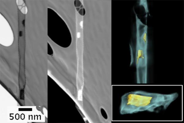
Fig. 2: (Left and Center) STEM Bright- and Dark- field compositional images of carbon tubes filled with Co nanoclusters - Beam energy 30 keV. – (Right) Tomogram of the cobalt clusters inside the carbon tubes. The inset shows one Co particle, demonstrating the chemical sensitivity and the monotonically variation of the contrast upon tilting. |