IT-2-P-3197 Temperature dependence of Z-contrast in InGaN
Scanning transmission electron microscopy (STEM) combined with a high angle annular dark field detector (HAADF) gives rise to image contrast strongly depending on the nuclear charges of the scattering specimen atoms and is often referred to as Z-Contrast. The comparison of HAADF-STEM image intensities with simulated intensities from multislice calculations allows determining specimen thickness or material composition for each atomic column [1, 2] in a high resolution HAADF-STEM micrograph.
The main contribution to the measured signal in HAADF-STEM stems from thermal diffuse scattering (TDS) due to the thermal vibrations of the specimen atoms. An increase of temperature should result in a higher HAADF-signal due to the larger amount of generated TDS.
In this contribution we have studied the thermal dependence of Z-contrast for InGaN/GaN and specimen temperatures between 300K and 600K. Fig. 1 shows the intensity profile of an five-fold InGaN/GaN multi-quantum well structure measured at different temperatures. We observed an increase of the HAADF-STEM intensity with increasing temperature for GaN as well as for InGaN. However, we also noticed that the material contrast in the image (ratio between intensities for InGaN and GaN) decreased with temperature (Fig. 2).
In order to understand this effect, multislice simulations were carried out in the frozen phonon approach for different specimen temperatures using the STEMsim program [3]. A temperature dependent parameterization of Debye-Waller-Factors derived from density functional theory calculations [4] was used. The simulated material contrast in dependence of specimen thickness is shown in Fig. 3 for an indium concentration of 30% and for three different temperatures. The contrast decreases with temperature as is it was found in the experiment and is related to the effect of static atomic displacements (SAD). SADs occur, if atoms with different covalent radii share the same crystal lattice or sublattice., which is the case for In and Ga in InGaN. A rise of temperature increases the contribution of thermal diffuse intensity, whose material contrast is smaller than that of Huang-scattering [5] caused by SADs. Thus, the material contrast decreases with increasing temperature.
[1] Rosenauer et al., Ultramicroscopy 109, 1171-1182 (2009)
[2] Rosenauer et al., Ultramicroscopy 111, 1316-1327 (2011)
[3] Rosenauer and Schowalter, Springer Proc. in Phys. 120, 169-172 (2007)
[4] Schowalter et al., Acta Cryst. A 65, 227-231 (2009)
[5] Z. L. Wang, Acta Cryst. A 51, 569-585 (1995)
This work was supported by the Deutsche Forschungsgemeinschaft under Contract No. RO 2057/8-1 and the Bundesministerium für Bildung und Forschung (BMBF) in the frame of the “ERA-SPOT True Green (13N9634)” project.

Fig. 1: HAADF-STEM intensity profile of a five-fold InGaN/GaN multi-quantum well structure grown on GaN measured at specimen temperatures of 300K, 450K and 600K. The intensity is increasing with increasing temperature. |
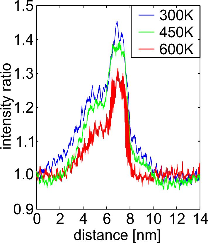
Fig. 2: Intensity profile of an InGaN quantum well normalized with respect to the intensity of the neighboring GaN for different specimen temperatures. |
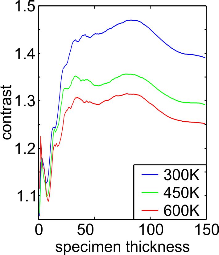
Fig. 3: Simulated material contrast (IInGaN/IGaN) of In0.3Ga0.7N for different specimen temperatures. |
|