IT-7-O-3133 In-situ Nano-compression tests on Shape Memory Alloys
Recently, there has been growing interest in the potential use of shape memory alloys (SMA) in micro and nano-scale structures and devices, for example as sensors or actuators in micro electromechanical systems (MEMS). With a growing worldwide market in excess of hundred billion dollars, MEMS constitute a new paradigm of technological development for the present century, and smart materials are converging with miniaturization technologies, enabling a new generation of smart MEMS or SMEMS. Among the different smart materials targeted for use in SMEMS, shape memory alloys (SMA) have attracted considerable interest because they offer the highest work output density, about 107 J/m3, and exhibit specific desirable thermo-mechanical effects such as superelasticity and shape memory.
In previous works, completely recoverable superelastic strain and shape memory in micro and nano pillars was first reported for Cu-Al-Ni SMAs [1] showing the competitive advantage of these SMAs over the commercially used of Ti-Ni. In addition several size effects on superelastic behaviour were also demonstrated [2, 3] in Cu-Al-Ni SMAs. For practical applications the superelastic behavior must be reproducible in order to be functionally reliable, and first studies on cycling SMA micropillars by nano compression tests were recently published [4, 5].
In this work we present an In-situ characterization of the nano-compression superelastic behaviour of Cu-Al-Ni micro-pillars at the scanning electron microscope. Micro-pillars were milled by Focused Ion Beam technique on [100] oriented Cu-Al-Ni single crystals. All pillars were tested in an instrumented pico-indenter Hysitron PI-85, introduced inside the chamber of a JEOL-FEG 7500, by using a diamond flat indenter, as can be seen on Figure 1. The nano-compression stage was tilted in order to allow imaging by the SEM. Simultaneous video-image was taken during the nano-compression test acquisition data in order to correlate the mechanical behaviour with microstructure evolution. Fully recoverable and reproducible superelastic behaviour has been obtained and a picture of the screen containing both, image and mechanical test, is shown in Figure 2.
[1] J. San Juan, M. L. Nó, and C. A. Schuh, Advanced Materials 20 (2008), p. 272.
[2] J. San Juan, M. L. Nó, and C. A. Schuh, Nature Nanotechnology 4 (2009), p. 415.
[3] J. San Juan and M. L. Nó, J. Alloys & Compounds 577S (2013), p S25.
[4] J. San Juan, M. L. Nó, and C. A. Schuh, Acta Materialia 60 (2012), p. 4093.
[5] J. San Juan, J. F. Gómez-Cortés, G. A. López, C. Jiao, and M. L. Nó, Appl. Phys. Lett. 104 (2014), p.011901
The authors thank the Spanish Ministry of Economy and Competitiveness, MINECO, project MAT2012-36421 and the CONSOLIDER-INGENIO CSD2009-00013, and the Basque Government for Consolidated Research Group IT-10-310 and ETORTEK-ACTIMAT-2013. J. San Juan and M.L. Nó also thank EOARD Grant FA8655-10-1-3074. J.F. Gómez-Cortés thanks the Ph.D. Grant from MINECO.
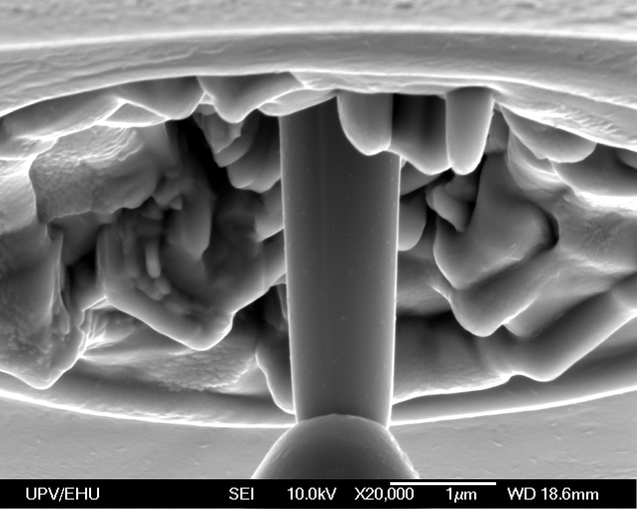
Fig. 1: Figure 1. Sub micrometre pillar of Cu-Al-Ni SMA, milled by focused ion beam, just before the in-situ nano-compression test. On the lower side of the image it can be appreciated the flat tip of the diamond indenter in contact with the top of the pillar. |
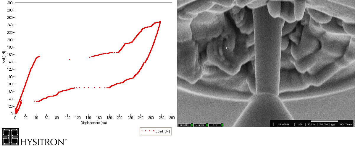
Fig. 2: Figure 2. In-situ superelastic nano-compression test performed on a sub-micrometre pillar of Cu-Al-Ni SMA. The video image allow to correlate the different points of the load-displacement curve with the corresponding images taken at the SEM. The above image corresponds to the final screen image just after finishing the in-situ test. |