IT-5-P-3124 Core/shell structure in magnetic nanoparticles from HRTEM and EELS
Magnetic properties on nanoscale materials are strongly affected by the possible different composition of the surface with respect to the bulk material.
In this study we investigate the composition and morphology of spherical d ≈ 10 nm magnetic particles, presenting a core/shell structure, synthesized through thermal decomposition of Fe-Co oleate under high boiling point solvents and posteriorly controlled surface oxidation. Core-shell structure has been assessed by X-ray diffraction analysis and corroborates the formation of two differentiated crystallographic phases: Co doped iron oxide spine (magnetite-like) and Co doped iron monoxide (wustite-like). From a careful inspection of HRTEM images (acquired on an aberration-corrected JEOL JEM-2200FS), an extra reflection from the shell region is indeed visible. This is compatible with a (220) reflection from magnetite. The (400) magnetite and (200) wustite reflections are indeed close (about 0.21 nm), indicating that some solubility or epitaxy of the two structures is possible.
HAADF images and spatially resolved EELS maps are acquired an aberration-corrected FEI Titan “cubed” microscope equipped with an electron monochromator and Gatan Enfinium. The core/shell structure of the particles is evident in HAADF. The reflection from magnetite is visible in the shell region. Moreover EELS maps, obtained by model-based fitting, reveals that there is a depletion of cobalt in the magnetite shell, together with the expected increasing in oxygen with respect to wustite. This is accompanied by the change in valence state of the transition metals from +2 to +3, as verified on Fe-L2,3 edge by fitting reference spectra. The energy position of the transition metal edge onset is indeed a valid parameter for determining its valence state.(1)
By further oxidation of the particles the core/shell structure seems to fade, the EELS maps revealing a more uniform distribution of Fe2+ and Fe3+ ions, indicating the magnetite phase extends towards the core. The final particles show higher policrystallinity with respect to the pristine particles. We can conclude that after further oxidation, the two phases are mixed in the whole volume. Further simulations from EELS profiles and/or EDS are in progress for quantifying the shell extension.
[1] H. Tan et al., Ultramicroscopy 116 (2012) 24–33
European Union FP7 Grant Agreement 312483 ESTEEM2 (Integrated Infrastructure Initiative–I3) and the European Union FP7 project 310516 NANOPYME
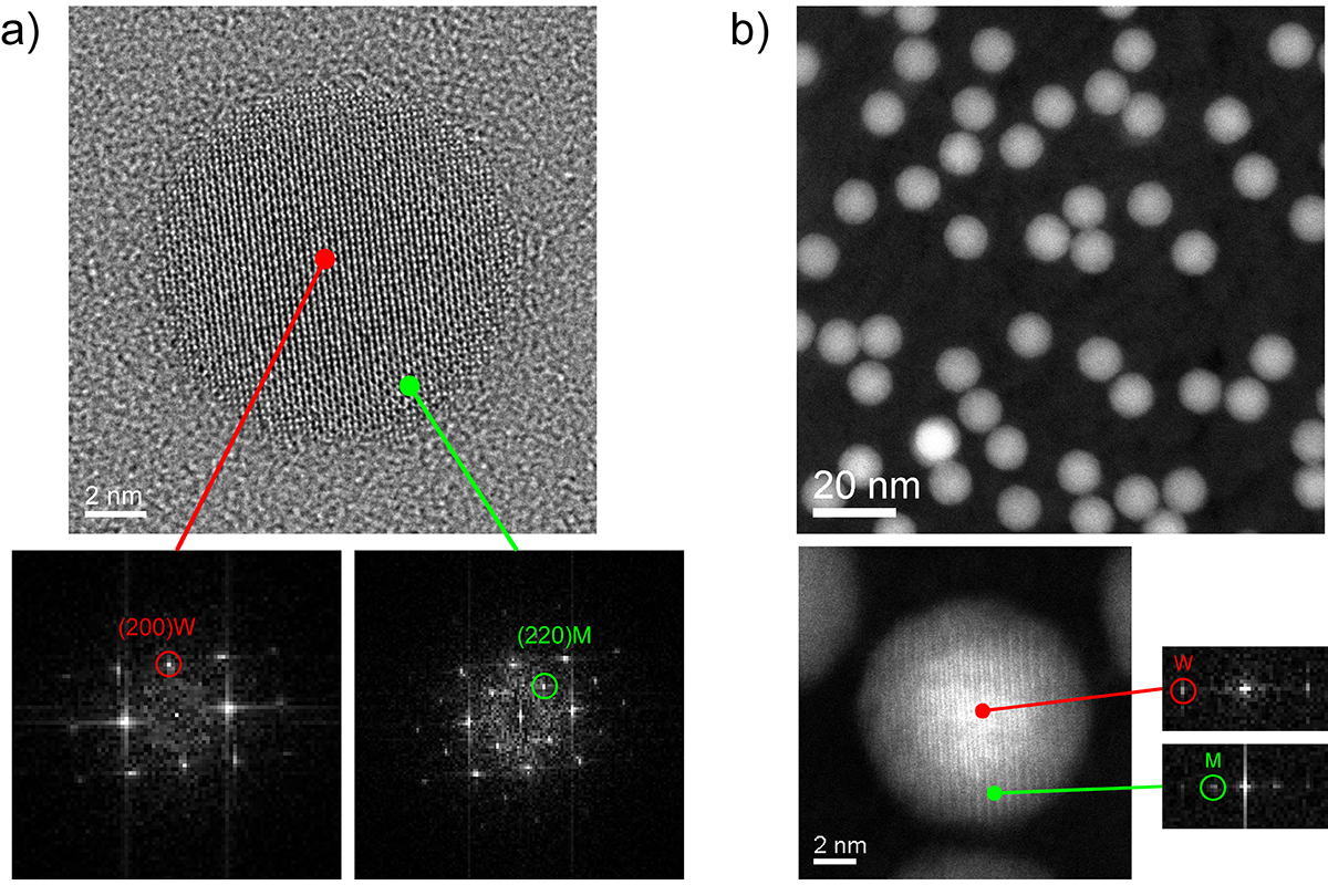
Fig. 1: a) HRTEM image from a Fe-Co oxide particle, showing an orientation close to [001] of a cubic structure. The FFT from the core can be addressed with wustite (W), while the shell region has clear reflections from (220) of magnetite (M). b) The core/shell structure as revealed in HAADF |
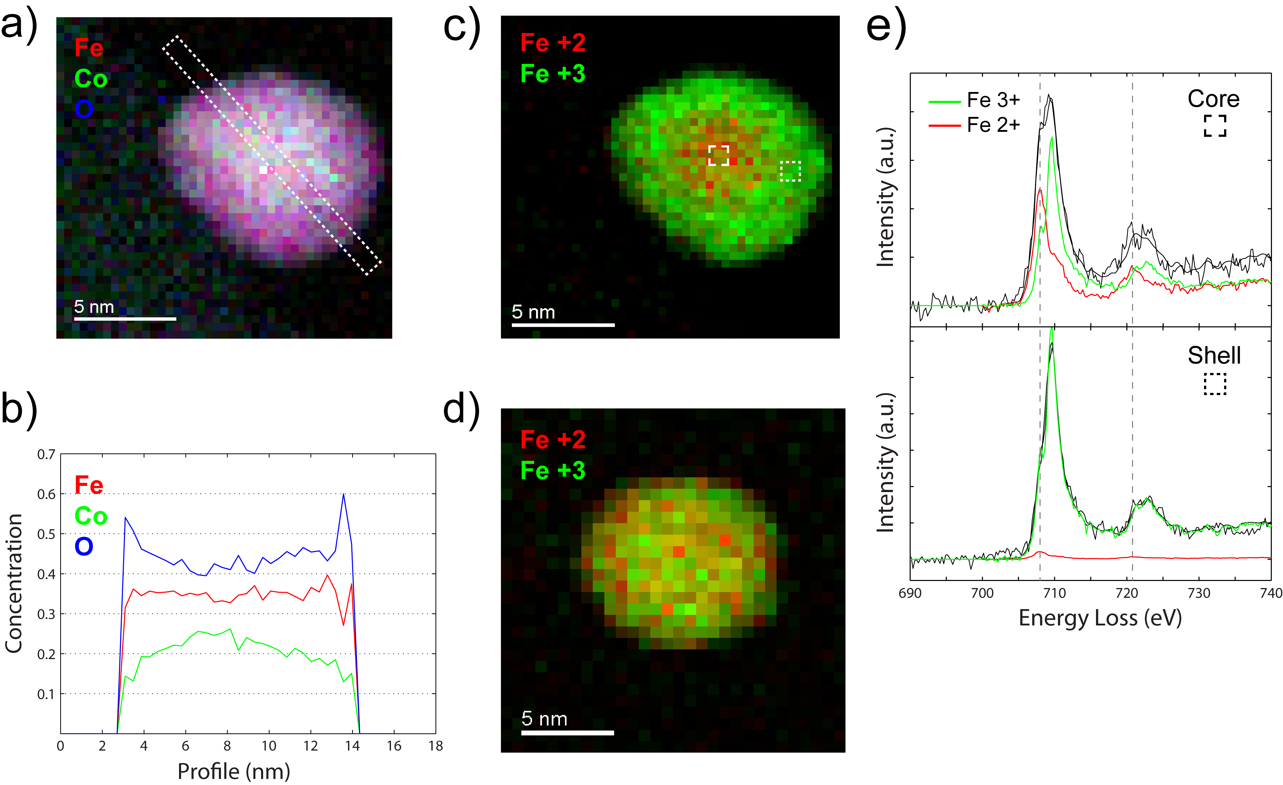
Fig. 2: a) Color map from model-based quantification; b) Linescan profile of the particle, showing the concentration of Fe, Co, and O, respectively; c) Valence map for Fe after fitting of reference spectra for Fe2+ and Fe3+ (explained in e); d) After further oxidation, there is no evidence of a core/shell structure |