IT-4-P-3090 Low-voltage STEM tomography: an alternative for soft polymers and hydrated samples
Tomography has become a key characterization tool in materials science as well as in biology. The principle of tomography is based on the acquisition of a series of projections images at different tilt angles, computation of the volume using dedicated algorithms and data segmentation and three-dimensional (3D) quantification. Several tomography techniques are available, using different types of radiations, depending on the observation scale. X-rays are currently used for the 0.5 µm–1 mm resolution level and the three-dimensional characterization of nanoscaled structures requires transmission electron microscopy (TEM) tomography or an atom-probe approach. At the mesoscopic scale, corresponding to a resolution level between 10 nm and 500 nm, Scanning Electron Microscopy (SEM)-based techniques – such as Focused ion Beam (FIB) or serial block face SEM – use a slice-and-view method to directly obtain slices of the materials volume.
Moreover, in Environmental SEM (ESEM), the presence of the gaseous environment and the control of the sample temperature have also permitted the imaging of nanoparticles in liquid with a nanometer resolution, through STEM-in-SEM observations [1]. Its main advantage lays in the fact that water condensation or evaporation can be finely tuned by varying the environmental pressure, which enables in situ hydration / dehydration experiments.
In the first part of this presentation, we will briefly present an alternative tomography technique for the 3D characterization of materials at the mesoscopic scale. This method, called low-voltage STEM tomography, consists in performing tilted tomography in a SEM (in the transmission mode, the so-called STEM-in-SEM mode), see Figure 1 [2]. The potentialities of low-voltage STEM tomography will be compared to that of other 3D techniques through the study of polyurethane films containing two different kinds of carbon nanotubes (see Figure 2).
In the second part of this presentation, we will present the possibility of observing the 3D structure of hydrated materials [3]. In particular, we will discuss the role of different experimental parameters such as the temperature and the electron dose received by the sample. Two examples will be used: a porous material and a latex suspension. Monte Carlo simulations will also be used to estimate the resolution which can be expected in both cases.
[1] A. Bogner et al., Ultramicroscopy 104 (2005), 290-301.
[2] P. Jornsanoh et al., Ultramicroscopy 111 (2011), 1247-1254.
[3] K. Masenelli-Varlot et al., Microscopy and Microanalysis, http://dx.doi.org/ 10.1017/S1431927614000105
The authors acknowledge the CLYM (Centre Lyonnais de Microscopie) for the access to the ESEM XL30FEG microscope, the Agence Nationale de la Recherche and the Institut Universitaire de France for financial support.
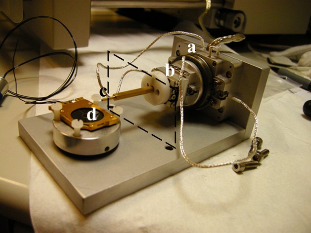
Fig. 1: Device for low-voltage STEM tomography, composed of a) and b) piezoelectric elements; c) sample holder and d) STEM detector. The dashed lines represent the position of the Peltier stage. |
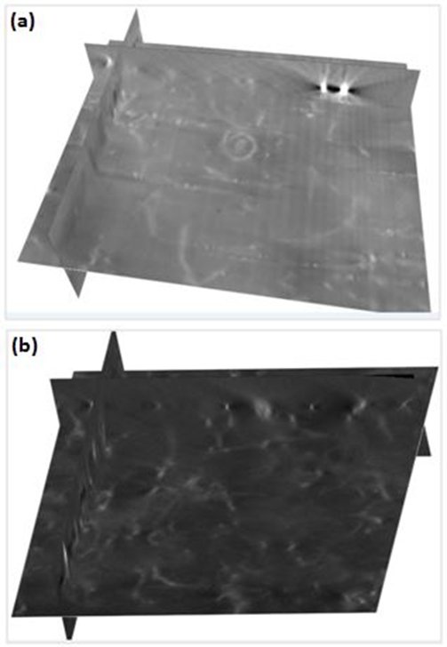
Fig. 2: Low-voltage STEM tomography on polyurethane thin films containing 2 vol.% of carbon nanotubes (CNT): orthogonal slices extracted from the volume. a) grafted CNTs and b) ungrafted CNTs. |