IT-5-P-3086 On the usability of electron vortices as probes for atomic resolution EMCD experiments
Recently discovered electron vortex beams, which carry a discrete orbital angular momentum (OAM) L, are predicted to reveal dichroic signals comparable to classical electron magnetic circular dichroism experiments (EMCD) [1]. Since electron beams can be easily focused down to sub-nanometer diameters, this novel technique provides the possibility to quantitatively determine local magnetic properties with unrivalled lateral resolution. For this purpose, specially designed apertures are needed to generate such non-planar electron waves [2]. Dichroic signals on the L2- and L3- edges are expected to be of the order of around 5% [3,4].
We have prepared and successfully implemented a spiral aperture into the condenser lens system of a FEI Titan3 80-300 transmission electron microscope (TEM) equipped with an image CS corrector (cf. fig. 1a). This setup allows for the generation of focused electron vortex beams with user-selectable OAM that can be used as probes in scanning TEM (STEM). Since for such spiral apertures, the different OAM are dispersed along the beam direction (z direction), the selection of the OAM is obtained by defocussing the beam.
First investigations aimed at probing the presence of an EMCD signal with such vortex beams were conducted on a 20 nm thin polycrystalline Ni film prepared by RF sputtering. Fig. 1b) shows the resulting EEL spectra subsequently acquired with L = +1 and L = -1 vortex states, respectively. In order to improve the signal-to-noise ratio, the binned-gain acquisition technique was used [5].
As can be seen from fig. 1b), these first experiments do not provide any evidence for differences in the absorption edges in the two EELS spectra.
In addition, the generation and propagation of the vortex wave functions and the spatial distributions of the OAM were simulated. The results of these simulations show that the orbital momenta and the beam intensity are largely localized (in all three dimensions) symmetrically around the geometrical focal points which are paraxial to the vortex cores (cf. fig. 2). Despite this localization, the superposition of contributions of vortex states (e.g., L = 0 and L = -1) adjacent on the one selected by appropriate defocussing (e.g., L = +1) are large enough that the average OAM is close to zero h, if the defocused portions of the wave are not properly prevented from interacting with the sample (cf. fig. 2b) which explains the absence of a dichroic signal in the experiment.
[1] J. Verbeeck et al., Nature 467 (2010), p. 301-304.
[2] J. Verbeeck et al., Ultramicroscopy 113 (2012), p. 83-87.
[3] P. Schattschneider et al., Ultramicroscopy 136 (2013), p. 81-85.
[4] J. Rusz and S. Bhowmick, Phys. Rev. Lett. 111 (2013), 105504.
[5] M. Bosman and V. J. Keast, Ultramicroscopy 108 (2008), p. 837-846.
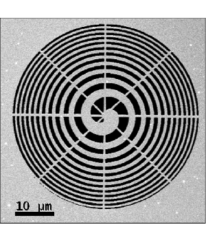
Fig. 1: SEM image of the spiral aperture installed in the C2 aperture plane of the electron microscope. |
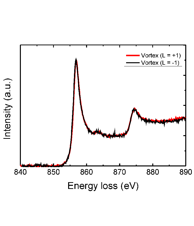
Fig. 2: Ni L3/L2 edges in the normalized EEL spectra acquired with electron probes of vortex states with orbital angular momenta L = +1 and L = -1, respectively. No significant EMCD signal (= difference between the two spectra) is observed. |
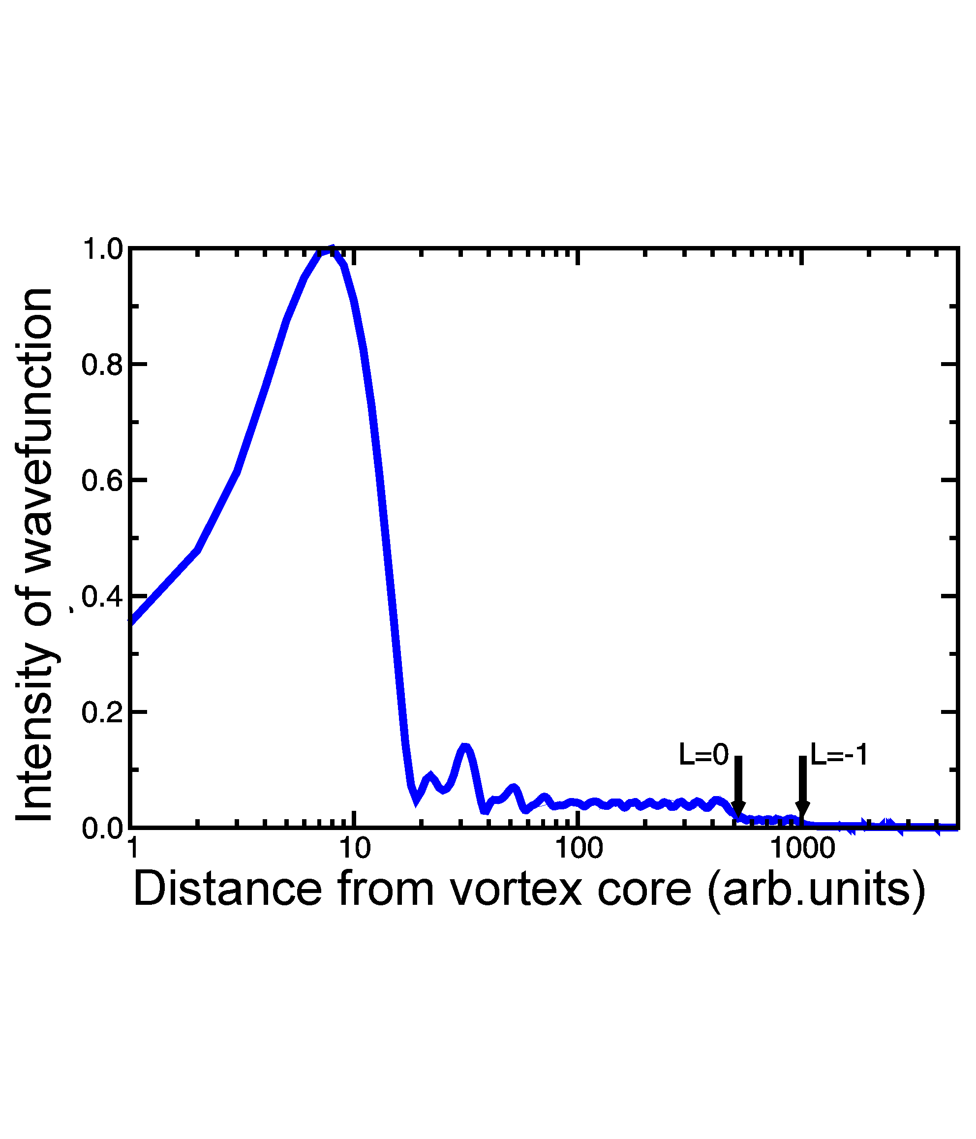
Fig. 3: Intensity profile of the simulated electron vortex beam with L = +1 generated with a spiral aperture. Arrows indicate radial positions where contributions of the L = 0 and L = -1 states become dominant. |
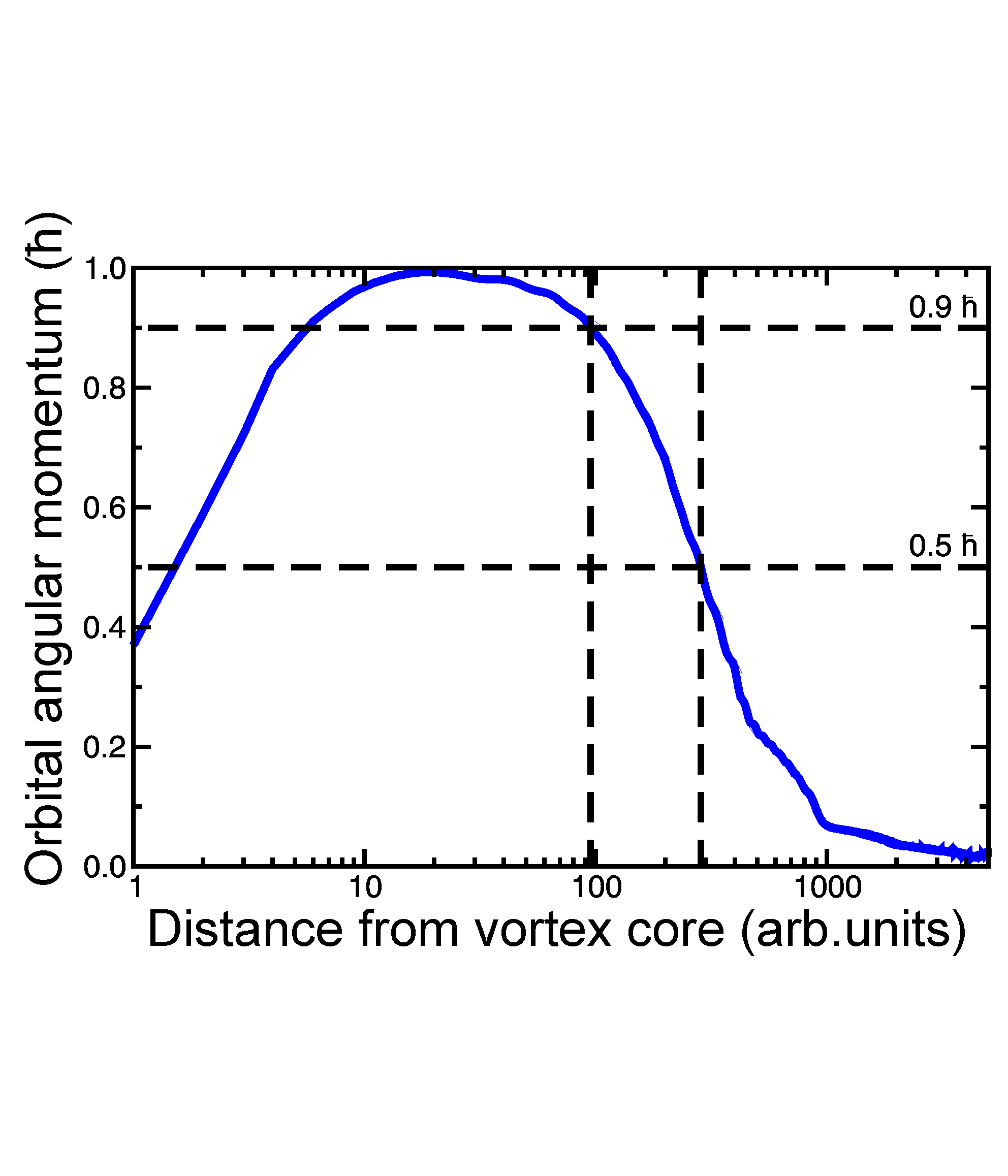
Fig. 4: Normalized expectation value of the OAM as obtained from radial integration from the vortex core to a given radius (1000 a.u. correspond to roughly 130nm). |