IT-7-O-3046 MAGNETIZATION REVERSAL PROCESS OF MAGNETIC SUPERDOMAIN STRUCTURES IN COBALT ANTIDOT ARRAYS
Geometric confinement of the magnetization in magnetic thin films by patterning regular antidot (hole) arrays has been considered a potential method to fabricate storage media of ultrahigh capacity or magnonic devices for high frequency applications [1]. Reducing the distance between antidots modifies favorably the magnetic properties towards the creation of individual magnetic entities that could be used as magnetic bit of information [2]. For this reason, it is necessary to use magnetic imaging techniques that can provide information of the local magnetic states at submicron scales. In this work, high spatial resolution Lorentz Microscopy (LM) combined with the in situ application of magnetic fields has been used to perform quantitative studies of the magnetic states of cobalt square antidot arrays with periodicities (p) ranging between 524 and 95 nm. Antidot arrays were patterned by Focused Ion Beam (FIB) etching on a 10-nm-thick cobalt film deposited on Si3N4 membrane. The FIB etching process produced holes of 55 nm diameter. At remanence, defocused LM images revealed a periodicity dependence of the magnetic domains and a transition in the domain wall geometry around p ~ 300 nm, changing from 90° and 180° walls to superdomain (SD) walls for small periodicities (see Fig. 1). A Fourier filtered method has been implemented to improve the direct visualization of one-dimension SD (magnetic chains) for arrays with p > 95 nm (see Fig. 2), which has allowed to determine the magnetic configuration inside the antidot cells in both the SD and the SD walls. The magnetization reversal processes by means of hysteresis cycles upon magnetic fields parallel and diagonal to the antidot rows have been studied by in situ LM experiments. As illustrated in Fig. 3, we have found that the reversal magnetization process occurs by simultaneous (parallel hysteresis cycle) or sequential (diagonal hysteresis cycle) nucleation and propagation of horizontal and vertical superdomain walls, respectively.
[1] Xiao Z L, Han C Y, Welp U, Wang H H, Willing G A, Vlasko-Vlasov V K, Kwok W K, Miller D J, Hiller J M, Cook R E and Crabtree G W 2003 Nanotechnology 3 357.
[2] Torres L, Lopéz-Diaz L and Iñiguez J 1998 Appl. Phys. Lett. 73 3766.
This work was supported by the Spanish Ministry of Economy and Innovation (MINECO) through the projects MAT2011-28532-C03-02 and MAT2011-23791 including FEDER funding, by the Aragón Regional Government through Projects E26 (MAGNA), E34 (IMANA) and CTPP4/11, and by the European Union under the Seventh Framework Program under a contract for an Integrated Infrastructure Initiative Ref 312483-ESTEEM2.
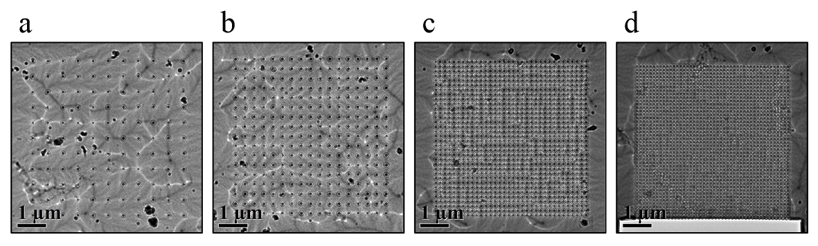
Fig. 1: Defocused LM images for square antidot arrays with periodicities of (a) 524 nm, (b) 327 nm, (c) 160 nm and (d) 116 nm. In the latter, magnetic contrast has nearly disappeared. |
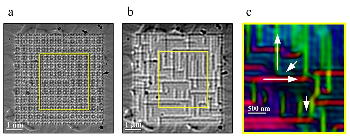
Fig. 2: (a) Raw and (b) Fourier filtered defocused LM images of the antidot array with p = 160 nm. (c) Color-coded magnetization orientation map obtained by the Transport-of-Intensity Equation reconstruction to a small region marked with a yellow rectangle in (a) and (b). |
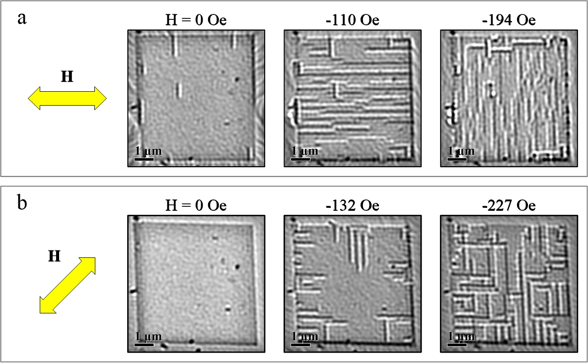
Fig. 3: Sequence of filtered defocus LM images recorded during the in situ application of in-plane magnetic fields (a) parallel and (b) perpendicular to the antidot rows. |
|