IT-9-P-3029 Strain Analysis by Nano-Beam Electron Diffraction (SANBED) in semiconductor nanostructures
The measurement of lattice strain is an important aspect in the characterisation of semiconductor nanostructures. As strain has large influence e.g. on the mobility of charge carriers, methods for accurate strain measurement with high precision are mandatory. In the present work [1] we measure strain from the positions of diffraction discs in convergent-beam electron diffraction (CBED) patterns using dedicated algorithms. Large one- and two-dimensional series of CBED-patterns (~3000) in semiconductor nanostructures have been recorded at an FEI Titan facility in STEM mode. Contrary to parallel illumination in conventional Nano-beam electron diffraction (NBED), we show that focusing the beam with a semi-convergence of 2.6 mrad increases the spatial resolution drastically by a factor of 5 to be 0.5 – 0.7nm. We determined the precision of this method to be 0.07%.
The rich inner structure of CBED-discs causes a big challenge to recognize their positions accurately. In particular, three different algorithms have been developed: As shown in figure 1, the first algorithm detects edges around each disc and iteratively deselects erroneous edges by circle-fitting. In this way the fit converges to the disc boundary. A disadvantage of this parameter-free method is a long computation time. An improvement of speed by a factor of 15 is achieved with the Radial Gradient Maximisation method. This method positions two sets of rings around the initially estimated disc position, one set of rings with smaller radii than estimated and one with larger radii, as illustrated in figure 2. Disc position and radius are determined by maximising the difference between the integrated intensity on inner and outer rings. The third method is a cross-correlation with different masks. As it is nearly a factor of 100 faster than the edge detection algorithm it is capable for in-situ strain measurement during CBED pattern acquisition. The left part of figure 3 shows two different masks. Mask A assumes fully illuminated CBED-discs, whereas with mask B the inner structure of the diffraction discs has less influence on the result. The right part of figure 3 shows the change in the correlation function if the disc is shifted. The disc position can be determined from the shift between mask and experiment. We compare the precision of the three different algorithms among each other and with respect to former approaches [e.g. 2, 3]. Finally we show by application, that specimen cooling, zero-loss energy filtering and aberrations of the projection system only weakly affect the measured strain.
[1] K. Müller and A. Rosenauer et al., Microsc. Microanal. 18 (2012), p. 995.
[2] F. Uesugi et al., Ultramicroscopy 111 (2011), p.995.
[3] A. Béché et al., Appl. Phys. Lett. 95 (2009), p. 123114.
This work was supported by the Deutsche Forschungsgemeinschaft (DFG) under contracts numer RO2057/8-1, SCHO1196/3, V0805/4 and V0805/5.

Fig. 1: Disc position and radius recognition with selective edge detection. (a) Process from experimental raw data to fitted disc position. (b) Edge point deselection: Beginning with the first circle-fit to all edge points, the point with the largest distance to the fitted circle is deleted until only edge points on the disc boundary remain. |

Fig. 2: Disc position and radius recognition with Radial Gradient Maximisation. Two sets of rings are positioned inside and outside an initial radius estimation and the intensity is integrated. Radius and position are fitted via maximisation of the difference between both sums. |
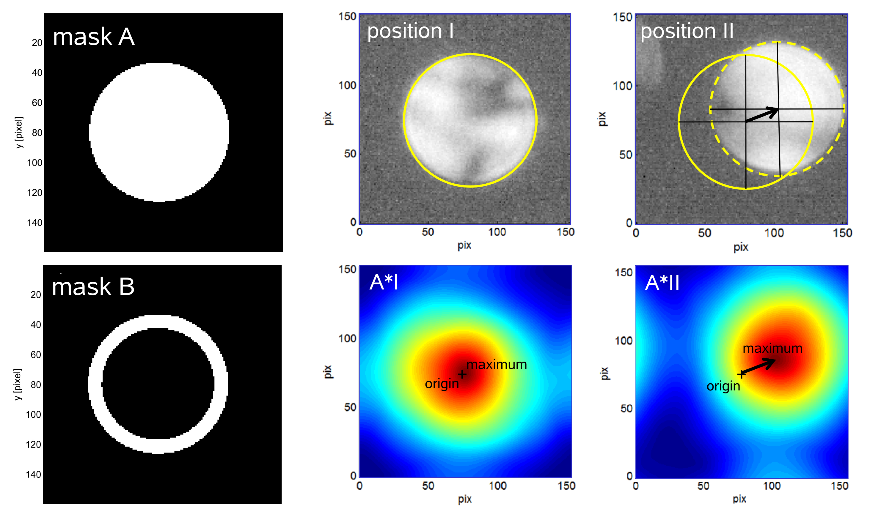
Fig. 3: Disc-position recognition via cross-correlation with masks. Left: Masks for correlation. Right: Changes in correlation-function (down) when the experimental disc shifts. Disc-position recognition from shift between experiment and mask. |
|