IT-8-O-3019 High Throughput Imaging in a Multibeam SEM
Nowadays there is a growing demand to increase the throughput of scanning electron microscopes, especially in biological research where 3-D images of organ’s structures are desired but too time-consuming. By adopting our Multi-Beam Scanning Electron Microscope (MBSEM) and proper stage moving strategy, the time for constructing a 3D image of 1mm3 brain can potentially be dramatically reduced from 200 days to 1 day. There is also a place for high throughput imaging in the semi-conductor industry where inspecting patterned wafers often asks the high resolution of the SEM, but the standard SEM is too slow.
The MBSEM which we have developed is based on a regular FEI Nova-Nano 200 SEM, but equipped with a novel multi-electron beam source module containing a MEMS fabricated aperture array that delivers a 14x14 array of focused beams with a resolution and current per beam comparable to a state of the art single beam SEM 1,2 .
A Secondary Electron (SE) imaging system, a Transmission Electron (TE) imaging system and a Backscatter Electron (BSE) imaging system have been designed for this MBSEM. The most challenging issue for these 3 imaging systems is how to separate and collect different signals belonging to corresponding beams, especially considering that the pitch of the primary beams on the sample is only 0.5~5 µm, and that the systems should work well for different landing energies and working distances.
Here we will present analysis and the simulation results of the SE and BSE detection systems, and recent experimental results of TE imaging. For the latter we have made use of the in-vacuum high resolution light microscope developed for in-situ correlative microscopy3 as shown in figure 1. We demonstrate that all beams arrive at the specimen in a regular grid (figure 2) and that each beam gives a focused image (figure 2). We will discuss the detection problems that arise when so much data can be detected simultaneously.
References:
1. A. Mohammadi-Gheidari, P.Kruit, Nuclear Instruments and Methods in Physics Research A 645 (2011) 60
2. A. Mohammadi-Gheidari, C. W. Hagen and P. Kruit, J. Vac. Sci. Technol. B28, (2010) 1071
3. A.C. Zonnevylle, R.F.C. van Tol, N. Liv, A.C. Narvaez, A.P.J. Effting, P. Kruit and J.P. Hogenboom; Journal of Microscopy, (2013) doi: 10.1111/jmi.12071.
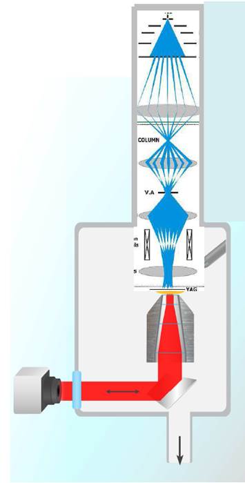
Fig. 1: Multi-Beam SEM with transmission detection |
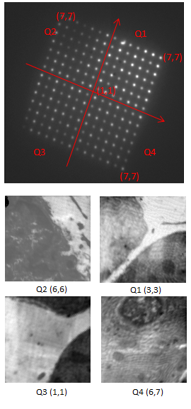
Fig. 2: Top: Multi-beam probes on the YAG : There are 14*14 beams in the system and the 4 quarters are used to identify beams. Bottom: TE images, based on the beam identification indicated above, TE images of different beams are shown, with the same field of view 4.3 µm, collected simultaneously. |