IT-11-P-3018 Experimental electron holographic tomography of magnetic vector fields in nanoscale materials
It is important to develop a characterization technique that can be used to measure three-dimensional magnetization distributions in nanoscale magnetic materials, which are of interest for a variety of applications that include future information storage technologies [1].
Off-axis electron holography allows the phase shift of the electron wave that has travelled through a thin sample to be measured in the transmission electron microscope (TEM) [2]. The phase shift is, in turn, sensitive to the in-plane component of the magnetic flux density within and around the specimen integrated in the electron beam direction. Although conventional tomographic reconstruction algorithms can in principle be used to reconstruct the magnetic flux density within and around a TEM specimen from two tilt series of magnetic phase images, significant artefacts can result from the difficulty of acquiring two ideal tilt series of images about independent axes over a complete range of specimen tilt angles.
We have therefore chosen to develop a different approach, which is based on the use of a model-based reconstruction algorithm to reconstruct the three-dimensional magnetization distribution in a TEM specimen, rather than the magnetic flux density, from two tilt series of phase images recorded using off-axis electron holography. Our approach involves repeated forward calculation of magnetic phase images until a best-fitting simulated magnetization distribution to the experimental images is obtained.
Practical challenges include the need to subtract the mean inner potential contribution to the phase shift recorded at each specimen tilt angle, the need to take into account the perturbed reference wave in the simulations, the minimization of contributions to the recorded phase from diffraction contrast and the preparation of a TEM specimen that is neither too thick nor obscured by another part of the specimen or the specimen holder when it is tilted to high angles about two axes.
Figure 1 shows simulated magnetic induction maps and magnetic phase images of an elliptical magnetic element for two different specimen tilt angles. Figure 2 illustrates the use of an electron-transparent silicon nitride membrane to acquire two independent tilt series of electron holograms of a lithographically patterned magnetic element. We will compare simulated and experimental results from both lithographically patterned elements and more three-dimensional nanoscale magnetic specimens and discuss the influence of noise and experimental artefacts on the final reconstructed magnetization distribution.
[1] S. S. P. Parkin et al., J. Appl. Phys. 85, 5828 (1999)
[2] D. Gabor, Proc. Roy. Soc. A 197, 454 (1949)
RDB acknowledges the European Commission for an Advanced Grant.
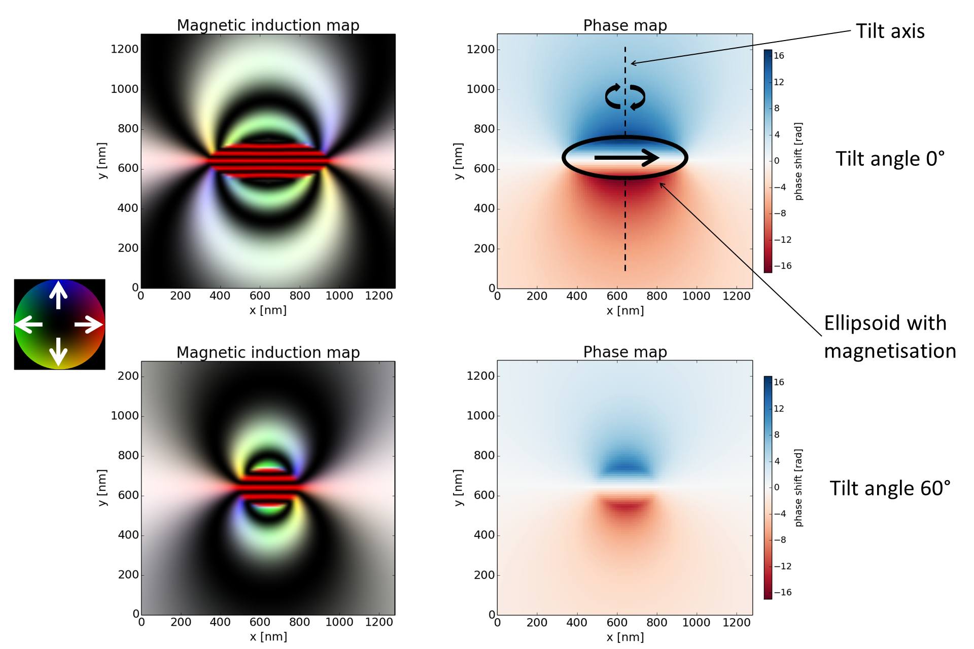
Fig. 1: Simulated magnetic phase shift and magnetic induction map of a uniformly magnetized elliptical element (saturation magnetisation = 3T) with semi-axis lengths of 600nm and 200nm and a thickness of 50nm for two different specimen tilt angles. The colors represent the direction and magnitude of the phase gradient, according to the color wheel shown. |
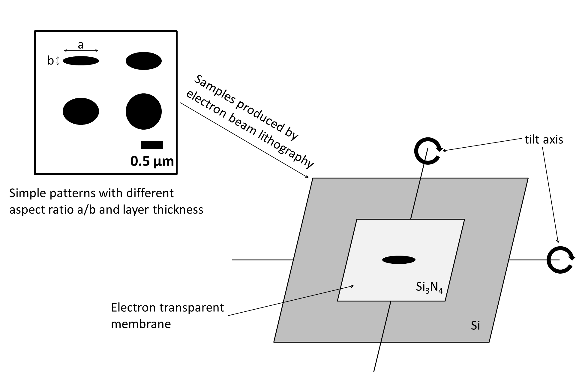
Fig. 2: Illustration of the design of lithographically patterned magnetic elements that can be tilted to high angles about two independent axes. |