IT-10-O-2962 Incoherent and coherent imaging for tomography of nano particle
Transmission electron microscopy (TEM) has been well recognized for its power in spatial resolution to the sub-Å level, especially, with aberration-corrected optics. However, the ultimate goal of electron microscopy is not only to obtain nice images but also to advance materials science. This means that EM has to evolve from describing to understanding materials properties. It is well-known that all the structure-property relations are encoded in the positions of the atoms and the shape of particle, specially, in the case of catalysts and biological species. The drawbacks of high resolution TEM are two folds. First, it gives only 2D projected structural information. And second, the passband of the lens transfer at the low spatial frequencies is very poor and such that the information about shape is lost.
In my talk, I will show that how we develop a novel theory and method for incoherent and coherent TEM imaging technique to determine 3D shape of nano-object with atomic resolution. For coherent imaging, our approach is to retrieve the three-dimensional atomic structure of nanocrystals from the electron exit wave function of a single projection image. The method employs wave propagation to determine the local exit surface of a sample together with the mass of each atomic column. Intensities are scaled by the mean inner potential of the sample and single atom sensitivity is expected since aberration-corrected electron microscopes are now available with such extraordinary capabilities. The validity of the approach is tested with a simulated exit wave function of a gold wedge, as shown in the fig. 1(a) and 1(b). The fig. 1(c) shows the reconstructed tomogram for the wedge Au crystal. For incoherent imaging, we present a new route to enhance the contrast by hollow cone imaging technique for biological objects using thermal diffuse scattered (TDS) electrons. Hollow cone imaging is incoherent and thus does not interfere with the central beam therefore it generates the amplitude contrast. Furthermore, the TDS signal is linear to the mass-thickness and easy to interpret and so it is suitable for soft material tomography. Here Fig. 2. we report the first results on the application of TDS to single particle analysis of proteins. The proof of the concept of the method has been demonstrated experimentally for Chaperonin GroEL as a standard protein since it is stable, easy to obtain and the structure is well-known.
CK is supported by the Office of Science, Office of Basic Energy Sciences of the U.S. Department of Energy under Contract No. DE-AC02—05CH11231. D. Van Dyck acknowledges the financial support from the Fund for Scientific Research - Flanders (FWO) under Project nos. VF04812N and G.0188.08 . F.-R. Chen would like to thank the support from NSC 96-2628-E-007-017-MY3 and NSC 101-2120-M-007-012-CC1.
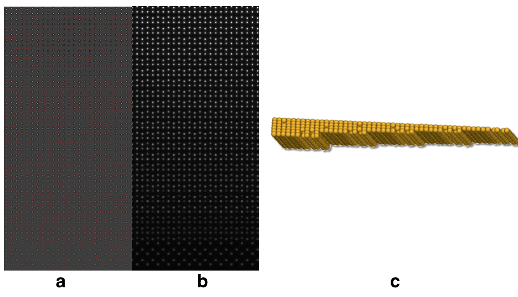
Fig. 1: Fig. 1(a) modulus of he simulated exit wave for Au wedge crystal (b) phase of simulated exit wave of Au wedge crystal (c) reconstructed tomogram from 1(a) and 1(b). |
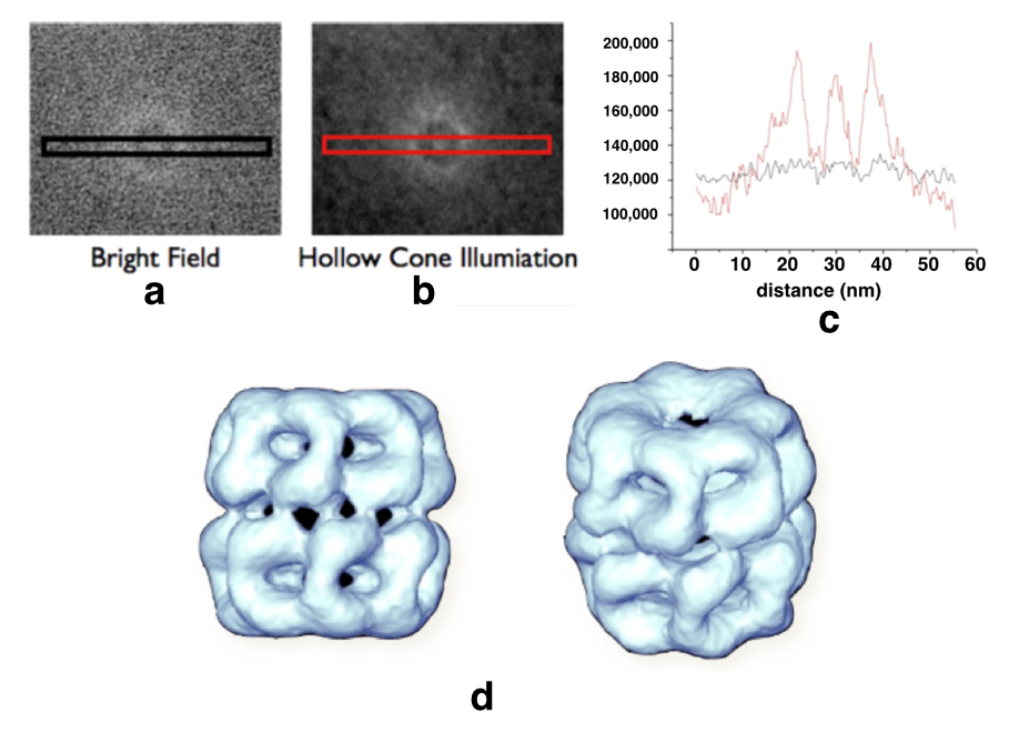
Fig. 2: Fig. 2 (a) Bright field image of GroEL (b) Hollow cone image (HCI) of GroEl (c) Intensity profile across the image in 2(a) and 2(b). (d) Reconstructed tomogram |