IT-1-O-2914 Tuning and Operation of a sub-20 meV Monochromator
When aiming for simultaneous high energy resolution and high spatial resolution in a monochromated scanning transmission electron microscope (STEM), three locations in the microscope are critical:
1) the monochromator’s (MC’s) energy-selecting slit, where the pass-band of energies admitted into the rest of the column is determined,
2) the sample, where the tuning determines the spatial resolution, and
3) the detector of the electron energy loss spectrometer (EELS).
To optimize the performance of the entire system, aberrations in all three locations must be accurately and repeatably tuned, so as to produce the smallest possible beam crossover at each place. In typical operation, all three crossovers are images of the field emission source, and upstream crossovers are re-imaged in subsequent stages. A mistuned monochromator can be largely compensated by a pre-sample aberration corrector that is mistuned in the opposite direction, or by a mistuned EELS.
The ideal method for monochromator tuning should therefore measure the actual aberrations at the plane of the energy selecting slit and not be affected by post-monochromator optics. We use a variation of the method developed by Foucault[1]: we image the far-field shadow of the energy-selecting slit near which the beam crossover is formed.
With a monochromatic beam coming into the monochromator, the aberrations would be tuned when the far-field image of the slit fades out uniformly as the slit is closed up. Non-zero focus and astigmatism would produce a stripe across the image of the beam-defining aperture, and one would focus and stigmate to make the stripe wider until it fills the aperture.
In practice, however, the slit is illuminated with an energy-dispersed beam some 300 meV wide, i.e. about 20 times larger than the energy width of our usual monochromated beam. This means that electrons with different incoming energies fill different parts of the aperture with stripes of different energies (Fig. 1), and the total beam after the slit is an incoherent superposition of a distribution of slit positions.
Fortunately, the tuning information is imprinted on the coherence properties of the beam exiting the MC slit, and we use this to determine the tuning at the slit to first and higher orders (Fig. 2). The end result is a repeatable tuning of the MC to 15 meV and better in Nion’s High Energy Resolution Monochromated EELS STEM (HERMESTM) (Fig. 3), as well as an ability to refocus the beam at the sample to sub-nm dimensions [2].
[1] L. Foucault, Comptes Rendus Academie des Sciences 47 (1858) 958-959.
[2] OL Krivanek et al, Microscopy 62 (2013) 3-21
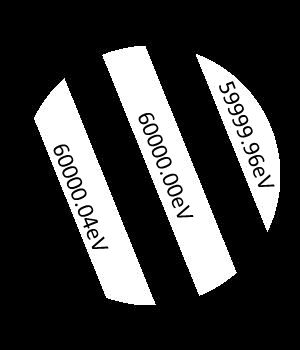
Fig. 1: Idealized far-field image (Ronchigram) for three electrons beams with slightly different energies, with astigmatism present at the MC slit. |
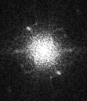
Fig. 2: Fourier transform of a Ronchigram obtained with the beam tuned at the MC slit. |
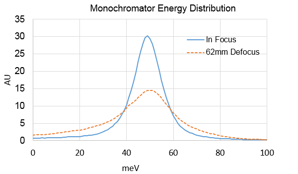
Fig. 3: Zero loss peaks before and after MC tuning |
|