IT-14-O-2891 Gaseous nanobubbles on immersed surfaces: Properties and imaging by AFM in situ and ex situ
Hydrophobic surfaces immersed in water are often densely occupied by gaseous nanodomains – nanobubbles and nanopancakes, with the size ranging between 10 – 100 nm, but it can exceed 1000 nm. The first direct proof of their existence came in the year 2001 in a form of in-situ atomic force microscopic (AFM) image made by J.W.G. Tyrrell and P. Attard [1] . Nevertheless, nanobubbles were still considered as artifacts, mostly due to their rather peculiar behavior, seeming disobeyance of Young Laplace law and hence nonexistence of plausible physical model. As various fields became affected by nanobubble existence, including interfacial physical chemistry, biophysics, microbiology, material sciences, nanofluidics, heterogeneous (electro) catalysis, immersion lithography and others, etc, nanobubbles started to attract growing attention. Their full impact is however, still to be disclosed.
Recent work performed in our laboratory [2] [3] [4] revealed the nanobubble ability to significantly rearrange solid surfaces which they adhere to. Nanobubbles can act as surface nanopatterning elements, changing in a significant manner its nanomorphology even at very mild conditions - in deionized water, at room temperature and pressure variations not exceeding 10 kPa. Besides nanobubble imaging by AFM in situ and distinguishing nanobubbles from solid nano-objects, we are presenting our novel technique, which can be called „nanobubblegraphy“[4], due to its ability to allow ex situ recognition and ex-post imaging of nanobubbles on dried surface as imprints developed after relatively short (sub-second) exposition in polymeric matrix (Figs 1 - 4).
Nanobubble properties and their utilization for imaging in situ and ex situ are discussed in relation to current physical models.
References
[1] J. W. G. Tyrrell and P. Attard: Phys. Rev. Lett. 87, 176104 (2001)
[2] P. Janda, H. O. Frank, Z. Bastl, M. Klementová, H. Tarábková, L. Kavan, Nanotechnology 21 (2010) 095707 (7pp)
[3] Viliam Kolivoška, Miroslav Gál, Magdaléna Hromadová, Štěpánka Lachmanová, Hana Tarábková, Pavel Janda, Lubomír Pospíšil, Andrea Morovská Turoňová: Colloids and Surfaces B: Biointerfaces 94 (2012) 213– 219
[4] H. Tarábková, P. Janda: J. Phys.: Condens. Matter 25 (2013) 184001
Acknowledged project support GACR P208/12/2429
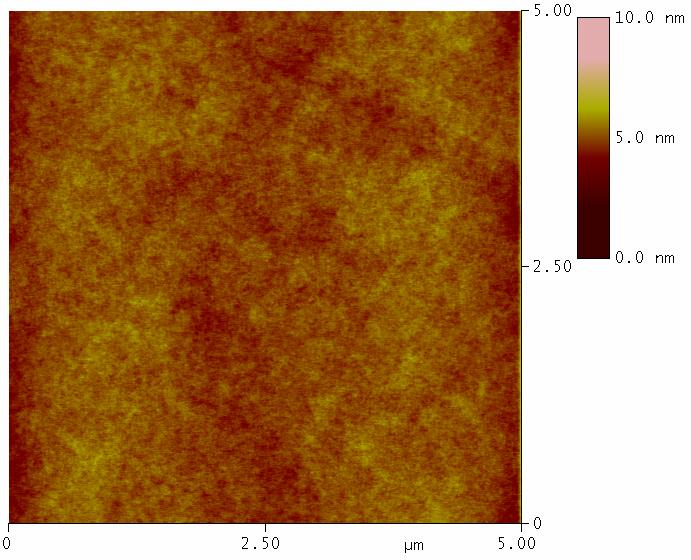
Fig. 1: AFM image of polystyrene film as received by spin-coating. |
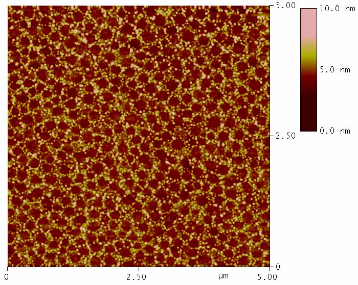
Fig. 2: AFM image of polystyrene film after exposition to nanobubbles in deionized water. Net-like nanopattern corresponds to 2D nanofoam imprinted into polystyrene matrix. |
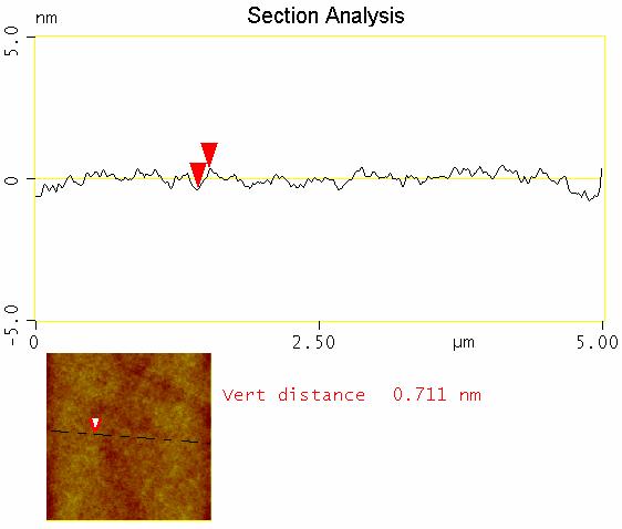
Fig. 3: Profile analysis of polystyrene film as received by spin coating (data from Fig. 1) |
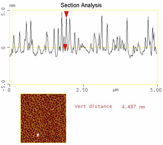
Fig. 4: Profile analysis of polystyrene film after exposition to nanobubbles in deionized water (data from Fig. 2) |