IT-7-P-2870 Revealing dislocation activities and deformation behavior in Nb2AlC using in situ nanoindentation in the transmission electron microscope
MAX phases are layered crystals with ternary or quaternary chemical composition. Due to their excellent electrical and thermal conductivity, as well as high oxidation resistance, they are in focus of intense research activities. Dislocation activities in MAX phases at room temperature (RT) are believed to be limited to slip along basal planes. In cyclic stress-strain curves, fully reversible, rate independent and closed hysteresis loops are observed. These features are attributed to the formation and annihilation of incipient kink bands (IKBs) and dislocation walls (DWs) [1]. Fig. 1 illustrates the formation of a KB. Initiated by elastic buckling above a critical maximum shear stress dislocation pairs of opposite sign form and move in opposite direction. It is believed that a nucleated KB immediately extends to the free surface. Hence, the attraction force between DWs disappears and a kink boundary is formed. An IKB does not dissociate into mobile DWs and thus is reversible, when the load is removed. KBs were observed ex situ by transmission electron microscopy (TEM) in single crystal Ti3SiC2, after nanoindentation perpendicular to basal planes (Fig. 1e) [2]. However, to date, the precise nucleation mechanism of IKBs and DWs is not known.
By means of in situ indentation in the TEM we reveal dislocation activities in Nb2AlC [3]. Undeformed Nb2AlC specimens exhibit basal plane dislocations with 1/3<11-20> type Burgers vectors. In situ indentation in dislocation-free regions and parallel to the basal planes (Fig. 3) reveals that basal plane dislocations nucleate and move in the same slip system without cross-slip or entanglement (Fig. 4). This confirms that these dislocations are mobile at RT, as proposed by Farber et al. in Ti3SiC2 [4]. The strain bursts in the load-displacement curve (Fig. 2) are assumed to be caused by dislocation nucleation. Indentation perpendicular to the basal planes results in an elastic deformation response, followed by fracture. Currently the formation mechanisms of IKBs and DWs are investigated in situ. Furthermore, plastic anisotropy is investigated by comparing pillar compression in the µm- and nm-range using scanning electron microscopy (SEM) and TEM, respectively. Prior to compression the pillar orientation is determined by electron backscatter diffraction with SEM and electron diffraction with TEM. For compression edge-on to the basal planes it is assumed that KB formation is more likely to occur than in pillar compression parallel to the c-axis.
[1] Barsoum et al., Nat. Mater. 2 (2003): 107
[2] Molina-Aldareguia et al., Scr. Mater. 49 (2003): 155
[3] Kabiri, Master Thesis, University Erlangen-Nuremberg (2013).
[4] Farber et al., J. Amer. Ceram. Soc. 81 (1998): 1677
[5] Barsoum et al., Metall. Mater. Trans. A 30 (1999): 1727
Financial support by the DFG via research training group GRK 1896 is gratefully acknowledged. The authors further thank Prof. Dr. Peter Greil for providing the samples.
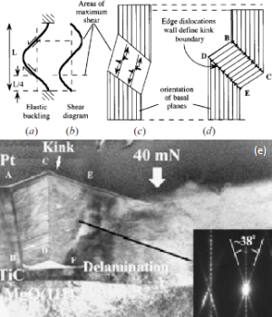
Fig. 1: Schematic of kink band formation: (a) Elastic buckling, (b) corresponding shear diagram, (c) formation of dislocation pairs and (d) kink band [5], (e) Cross-sectional TEM image of an indent in a Ti3SiC2 (0001) single-crystal thin film with a maximum load of 40 mN [2]. |
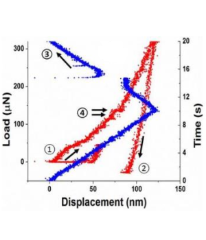
Fig. 2: Load-displacement and time-displacement curve of an in situ indentation parallel to the basal planes. Start and finish of the indentation corresponds to points 1 and 2 or 3, respectively [3]. |
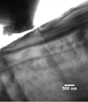
Fig. 3: TEM image showing the sample tilted in two beam condition around the <0001> zone axis, before an in situ indentation parallel to the basal planes [3]. |
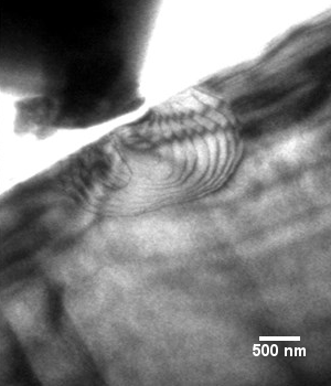
Fig. 4: TEM image after an in situ indentation parallel to the basal planes revealing nucleation and propagation of basal plane dislocations [3]. |