IT-12-IN-2866 Addressing Fundamental Problems in Information technology: Opportunities for X-Ray Photoelectron Spectromicroscopy
Modern information technology must exploit the full potential of complex material systems for the meticulous control of state variables. These state variables are used to encode an information bit and may be electron charges in semiconductor nanoelectronics, electron spins in the case spintronics, or local redox configurations in resistive switching elements. Consequently, the materials encompass intermetallic compounds, oxides or chalcogenides, elementary and compound semiconductors or even molecular components. In addition, the functional elements, for example, individual memory cells or transistor structures often involve nanometric dimensions and operate on nanosecond timescales or even below. This imposes considerable challenges on the characterization of electronic, chemical and magnetic states in the steady state or during operation.
Immersion lens microscopy with synchrotron radiation has matured into a versatile and powerful tool to investigate a broad range of issues in condensed matter physics and materials science. It combines high-resolution imaging with spectroscopic capabilities in a unique fashion. The excitation with photons from the soft to the hard x-ray regime ensures element selectivity and variable information depth. The polarization state of the synchrotron radiation enables a distinction of different magnetic orderings (Fig. 1), whereas the intrinsic time structure of the synchrotron radiation permits the study of processes with picosecond time-resolution.
In this contribution we will review the present status of x-ray photoemission spectromicroscopy with emphasis on applications in information technology. In particular, we will cover model systems in spintronics and in resistive switching (Fig. 2). The results will cover both static properties and dynamic processes. We will also discuss new developments, such as photoemission microscopy with hard x-rays and imaging spin polarimetry.
I would like to thank N. Barrett, S. Cramm, R. Dittmann, W. Drube, M. Escher, V. Feyer, A. Gloskovskii, A. Kaiser, J. Kirschner, A. Koehl, I. Krug, Ch. Lenser, M. Merkel, M. Patt, L. Plucinski, J. Rault, O. Renault, Ch. Tusche, N. Weber, R. Waser, and C. Wiemann for their cooperation. Financial support through the Deutsche Forschungsgemeinschaft (SFB 917) is gratefully acknowledged.
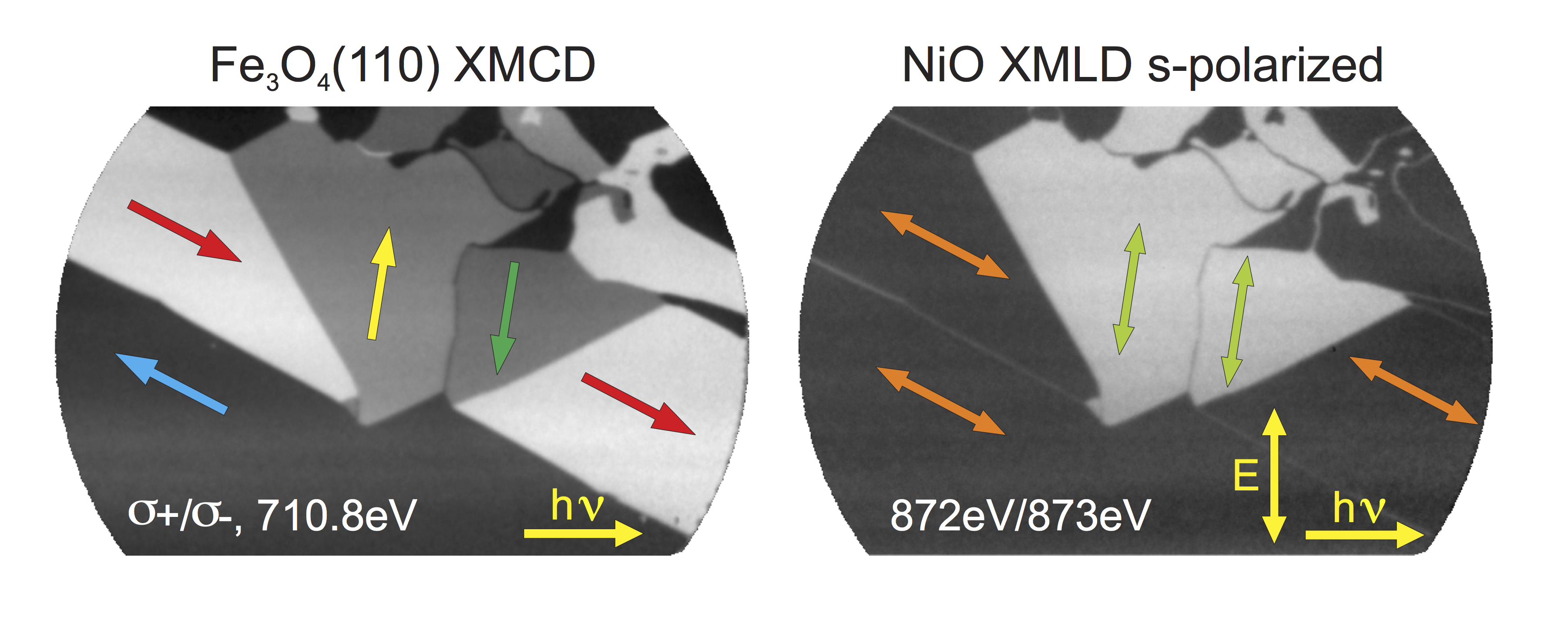
Fig. 1: Ferro- (left) and antiferromagnetic domain pat- terns in the system NiO/Fe3O4(110) exploiting XMCD (Fe) and XMLD contrast (Ni) at the indicated photon energies. Wide arrows indicate the local spin alignment axis. |
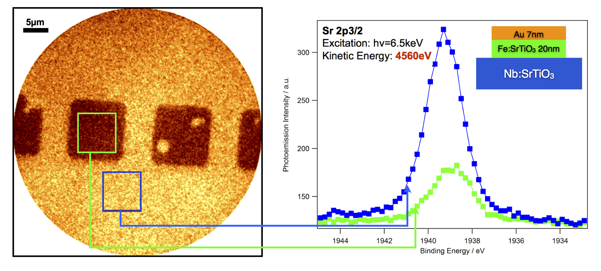
Fig. 2: Spatially resolved hard x-ray photoemission from a Fe:SrTiO3. The dark squares result from 7nm thick Au electrode pads deposited for the resistive switching experiments. |