IT-7-P-2853 Correlating internal structure and mechanical properties of amorphous silica and gold micro-/nanoparticles using in situ mechanical testing in the scanning and transmission electron microscope
The ongoing miniaturization and reliability of functional materials and devices at micro- and nanoscale inevitably necessitates information on small scale mechanical properties. In the fields of e.g. optics and biomedical imaging, nanospheres of both, silica and gold, as well as hybrid core-shell structures made of these materials are of great relevance [1,2]. In order to elucidate deformation mechanisms and related mechanical properties on the nanoscale, compression of individual particles is particularly suitable due to the absence of strain gradient plasticity effects [3].
In the present work we use in situ mechanical testing in the scanning electron microscope (SEM) and transmission electron microscope (TEM) with the aim to characterize the mechanical properties of amorphous silica and gold micro-/nanoparticles. Thereby a custom built SEM supported manipulation device [4] and a TEM Picoindenter® (Hysitron, Inc.) are used.
Silica spheres are synthesized according to the Stöber-Fink-Bohn method [5] followed by thermal treatments at 400°C (S400), 800°C (S800) and 1000°C (S1000), respectively. Structural changes and mechanical properties are studied using in situ SEM indentation (Fig. 1b), combined with solid state nuclear resonance spectroscopy (NMR) and infrared spectroscopy. Reduced Young’s modulus and hardness of untreated silica particles (S70) (29.6±6.2 GPa and 6.0±0.6 GPa, respectively) increase after calcination at 1000°C (58.2±8.8 GPa and 13.4±0.9 GPa, respectively), as shown in Fig. 2b. The values achieved after calcination at 1000°C agree well with values known for bulk fused silica. In the course of heat treatment, infrared and NMR spectra (Fig. 3) reveal condensation of internal OH-groups and enhanced cross-linking of the silica network, resulting in particles with chemically and mechanically similar properties when compared to their bulk counterpart [6].
Gold nanoparticles (20 nm – 1 µm in size) are prepared by solid/liquid state dewetting of thin gold films. In situ SEM compression experiments show large strain bursts after elastic loading. Currently, cross-sections of as-prepared and deformed Au particles are investigated by ex situ TEM to get more insights into the internal particle structure. Nanomechanical testing in the TEM is focused on smaller Au particles with the aim to reveal deformation mechanisms in situ.
[1] Oldenburg et al., Chem. Phys. Lett. 288 (1998):243.
[2] Fuller et al., Biomaterials 29 (2008):1526.
[3] Gao et al., J. Mech. Phys. Solids 47 (1999):123.
[4] Romeis et al., Rev. Sci. Instrum. 83 (2012):095105.
[5] Stöber et al., J. Colloid. Interface Sci. 26 (1986):62.
[6] Romeis et al., Part. Part. Syst. Charact. (2014).
The German Science Foundation is gratefully acknowledged for financial support within the priority program “Particles in Contact” and the research training group 1896.
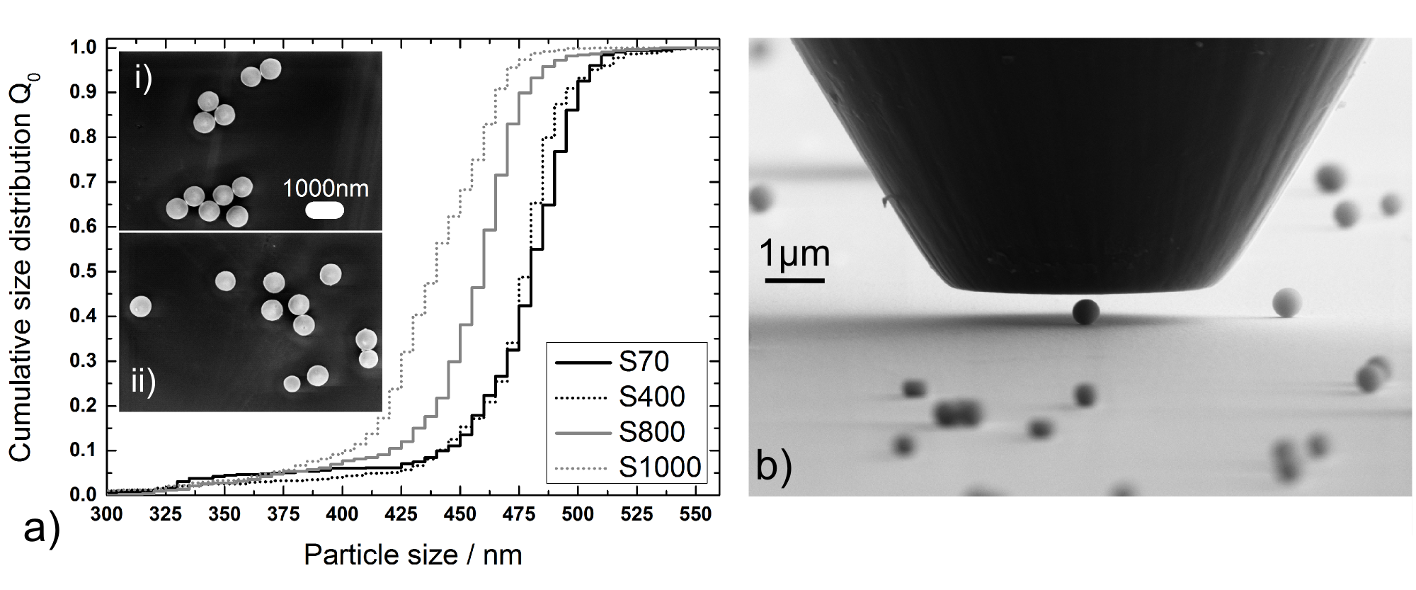
Fig. 1: a) Cumulative particle size distribution for untreated (S70) and heat treated (S400, S800, S1000) silica particles. Insets show representative SEM images for i) S70 and ii) S1000. b) Experimental setup for compression of single silica spheres between a diamond flat punch and a silicon substrate inside a SEM. |
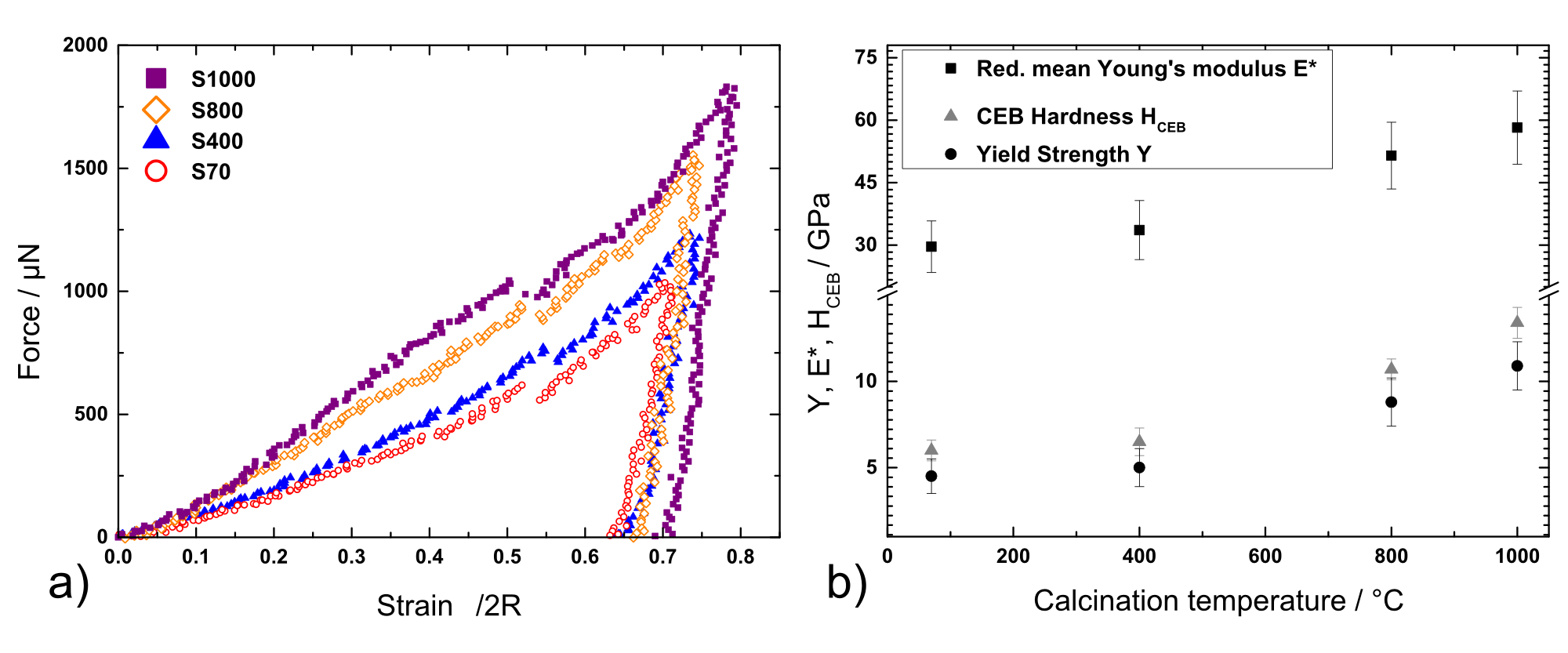
Fig. 2: a) Representative force-strain curves for silica particles from samples S70, S400, S800 and S1000. b) Reduced Young’s modulus, hardness and yield strength with respect to calcination temperature. At 1000°C, E* and HCEB approach the bulk values of fused silica. |
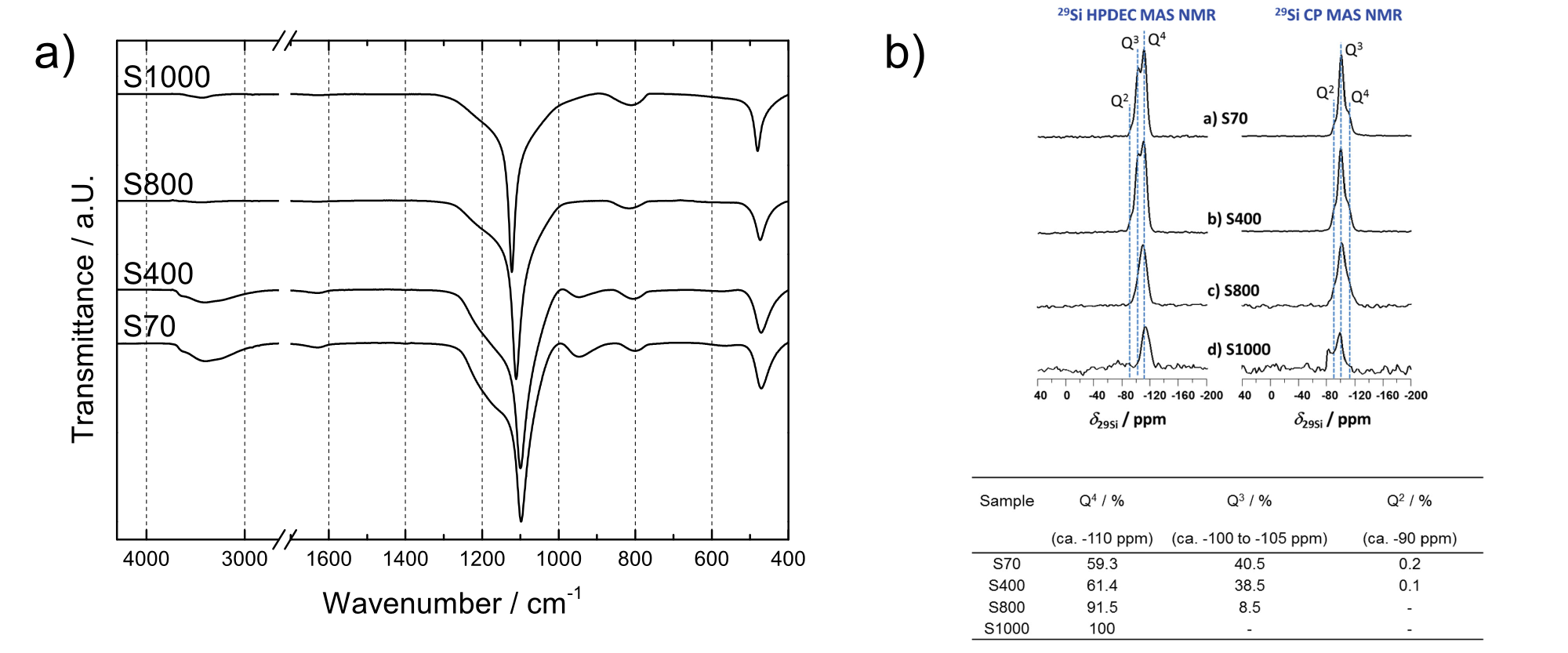
Fig. 3: a) Infrared spectra of the heated silica particles. For higher temperatures densification and dehydroxylation occur. b) 29Si HPDEC and 29Si CP MAS NMR spectra of the samples. The mean number of siloxane (Si-O-Si) bonds per silicon atom (Qi, 1≤i≤4) increases from S70 to S1000, confirming enhanced cross-linking of the silica network. |
|