IT-10-O-2806 Electron cryo-tomography with a new type of phase plate
Recent years have shown an increased interest in the development and use of phase plates in cryo-EM. The oldest and the most productive type of phase plate is the carbon film Zernike phase plate. Despite its good performance the Zernike phase plate has a few pitfalls. One major practical hindrance is its short lifetime, typically about 10 days. Another disadvantage is the generation of fringes around high-contrast features in the image. Despite its shortcomings the Zernike phase plate has been the main motivation and experience generator in the last years.
We are currently working in collaboration with FEI on the development and testing of a new type of phase plate. It addresses both of the above mentioned shortcomings of the Zernike phase plate. Our tests indicate that the new phase plate lasts for more than six months inside the microscope. This is a big advantage in terms of servicing and up time of the microscope. Another big advantage of the new phase plate is that it produces fringe-free images which resemble in appearance light microscopy phase contrast images. The new phase plate is being developed as a part of a product package which will include new hardware – phase plate holder & phase plate, and new software for alignment, calibration and ease of use.
We tested the new phase plate with two automated acquisition software packages – FEI Tomography and SerialEM. Both packages work well and are able to automatically acquire tilt series with the phase plate. There are only a couple of additional steps that are required for setting up the phase plate before starting the tilt series acquisition. The reconstruction process for the phase plate tomograms is almost identical to that for conventional defocus phase contrast tomograms and can be performed with any existing reconstruction software package, such as IMOD. Because of the strong low frequency components in the phase plate images a simple weighted back-projection reconstruction was in most cases sufficient to produce sharp, high-contrast tomograms. No further processing, such as de-noising, was necessary. In a few thick specimen cases a SIRT reconstruction produced better looking tomograms and again no de-noising or other post-processing was necessary. Overall the new phase plate works very well for cryo-tomography and users with cryo-tomography experience require only a short training on how to use it.
The new phase plate was tested in two microscope models – a 200 kV FEI Tecnai F20 and a 300 kV FEI Titan Krios. The lifetime and image quality performance was equally good with both microscopes.
We thank Matthijn Vos from FEI for providing test samples.
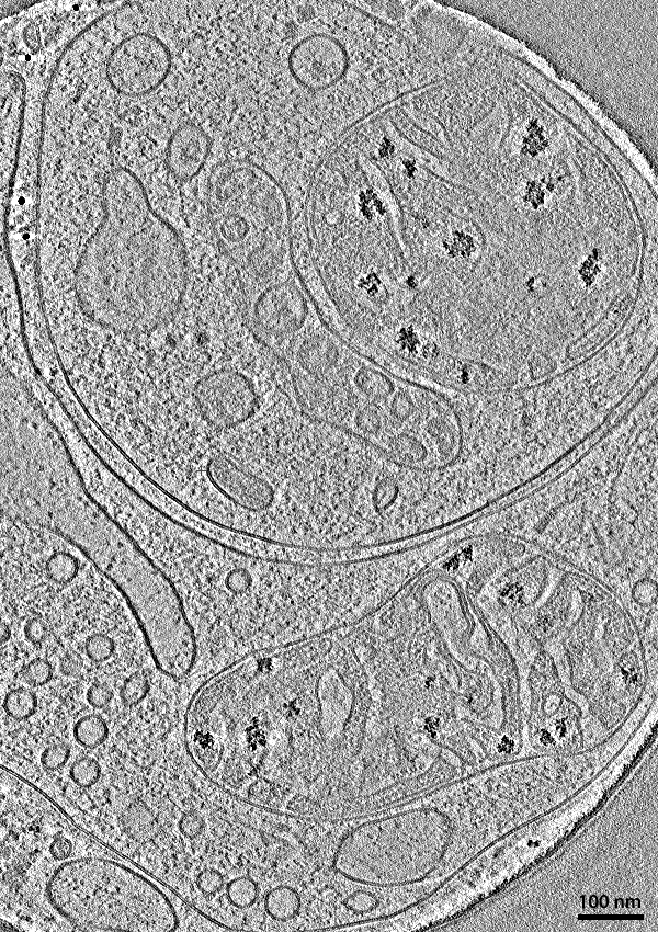
Fig. 1: A slice through a phase plate cryo-tomogram of a vitrified primary culture neuron cell. |
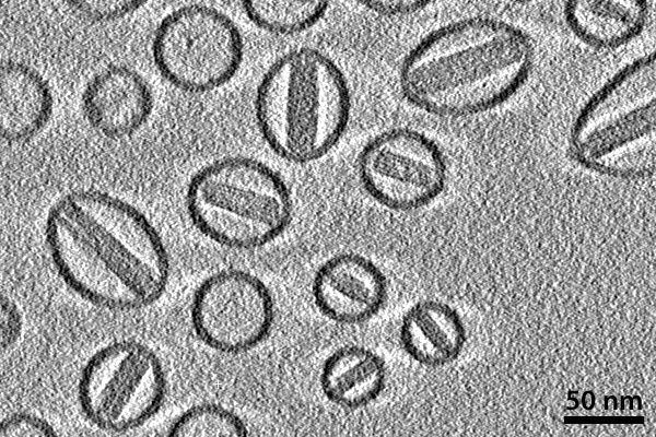
Fig. 2:
|