IT-8-P-2678 Implementing in situ experiments in liquids in the(scanning) transmission electron microscope and dynamic TEM
Developing a fundamental understanding of phenomena that take place in liquids, such as nanoparticle growth, protein conformational dynamics or the transformation of active materials during battery operation requires characterization tools able to provide in situ information with nanometer spatial resolution. In principle, this can be achieved using fluid stages in the (scanning) transmission electron microscope ((S)TEM). One of the main experimental challenges in the field is obtaining reproducible data free of beam-induced effects to enable quantitative analysis. Methods of calibration of the amount of radiation damage resulting from beam-induced reactions with the sample continue to be needed [1,2]. For instance, in situ growth of particles in solution by the electron beam is typically observed in (S)TEM experiments and has been used to calibrate the effect of electron dose in a Ag precursor solution in an in situ fluid stage [3] (Figure 1 (a) shows areas where Ag was grown under different experimental conditions). Custom image analysis algorithms can be applied to analyze movies of nanocrystal nucleation and growth and extract important information on growth dynamics and parameters such as the induction threshold below which no nucleation occurs [3] (see an example of image analysis in Figure 1(b)). Besides electron dose, factors such as accelerating voltage, imaging mode (e.g. TEM, STEM, SEM), liquid thickness, and solution composition are expected to affect the results of in situ experiments. Reproducing an experiment in a different instrument operating with different electron optical settings, introduces a large set of variables whose effect must be calibrated. Here, we present our recent developments in the design and implementation of calibration experiments using in situ fluid stages, including an identification of beam-sample interactions for changing imaging and experimental conditions. Since fluid stages are designed to fit in any transmission electron microscopy, the different capabilities of each instrument can be applied to the study of liquid phase reactions. When using fluid stages in combination with the dynamic TEM (DTEM), a combined temporal and spatial resolution of ~10-6 and ~10-10 m, could be achieved (see schematic of the DTEM design at the Pacific Northwest National Laboratory (PNNL) in Fig. 2). The unique qualities of the DTEM that benefit the in-situ experiments with fluid environmental cells will be also discussed.
[1] T.J. Woehl et al., Ultramicroscopy 127 (2013) 2927
[2]J.M. Grogan, Nano Letters 14 (2014) 359
[3] T.J. Woehl et al., ACS Nano 6 (2012), p. 8599; Nano Letters 14 (2014), p. 373.
[4] J.E. Evans et al., Microscopy 62 (2013) p. 147-156
This work was supported by the CII; under the LDRD Program at PNNL. PNNL is a multiprogram national laboratory operated by Battelle for the U.S. DOE under Contract DE-AC05-76RL01830. A portion of the research was performed using the EMSL, a national scientific user facility sponsored by the DOE's Office of BER and located at PNNL.
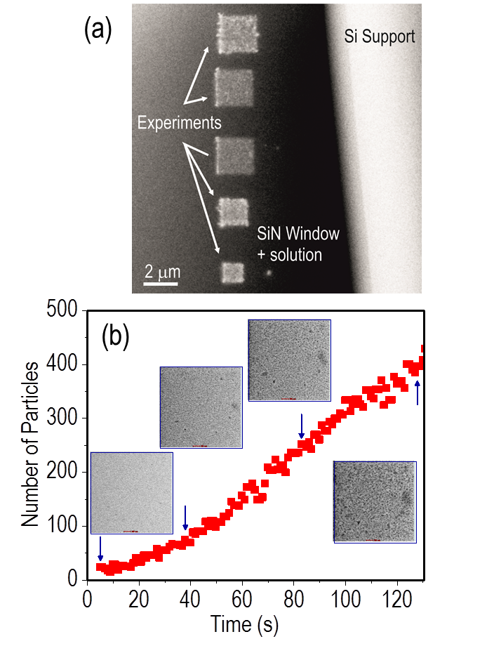
Fig. 1: (a) Low magnification BF STEM image showing a set of nanocrystals growth experiments using different electron dose rates. (b) Number of particles grown from solution as a function of time measured from an in situ dataset using 300kV, 7.1pA beamcurrent, 3 μs pixel-dwell time and M=40000x in STEM, to give a dose per frame of 39.1 e-/nm2f. |
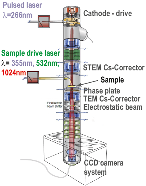
Fig. 2: Schematic of the DTEM showing theupgrades planed at PNNL. Modified from [4]. Copyright 2013 Oxford UniversityPress. |