IT-11-O-2667 Caustics and diffraction from two oppositely biased metallic tips imaged in the coherent transmission electron microscope
The coherence of a modern field emission transmission electron microscope (TEM) allows fascinating electron-optical phenomena to be observed, such as the fine structure of umbilic foci outlining the caustic of an astigmatic probe, the hyperbolic umbilic catastrophe produced by a coma aberration function [1] and cusped fan-like structures in defocused images of electrically biased nanotube bundles [2].
Here, we study bright-field TEM images of two oppositely-biased metallic tips, which show a rich structure that depends sensitively on applied bias and defocus as a result of a combination of electrostatic field topography and electron-optical phase shift and is strongly reminiscent of the elliptic umbilic diffraction catastrophe that occurs when visible light is refracted by a water droplet with a triangular perimeter [3].
An FEI Titan 60-300 field emission gun TEM was used to study two metallic tips that had been thinned electrochemically and mounted in a specimen holder equipped with piezo-electric drivers and electrical contacts. The tips were placed in front of each other at a separation of ~1 micron and a potential difference of up to 130 V was applied between them. The positively charged wire was found to act like a terminating convergent electron biprism, producing an overlapping region of intensity containing two-beam fringes, whereas the negatively charged wire acted like a terminating divergent biprism. The combined effect of the fields resulted in a highly complex interference pattern, which is shown in Fig. 1 for a nominal defocus of 9.5 mm and a potential difference of 130 V. The overlapping region has a triangular structure that is similar to the elliptic umbilic diffraction catastrophe. Figure 2 shows this region at a higher magnification, with the hexagonal structure of the spots and their modulation by the two-beam biprism fringes visible.
We have interpreted the key features in these images by using a simple model of two uniformly and oppositely charged lines placed in front of each other [4]. The shapes of the tips can then be approximated by suitably choosing two of the equipotential ellipsoidal surfaces. The resulting simulations shown in Figs 3 and 4 are in good qualitative agreement with the experimental results.
References
[1] T. C. Petersen et. al., Phys. Rev. Lett. 110 (2013) 033901.
[2] M. Beleggia et. al., Appl. Phys. Lett. 98 (2011) 243101.
[3] M. V. Berry, J. F. Nye, and F. J. Wright, Phil. Trans. Roy. Soc. London, Series A, 291 (1979) 453.
[4] M. Muccini et .al., Ultramicroscopy 45 (1992) 77.
We are grateful to C. Dwyer for valuable discussions.
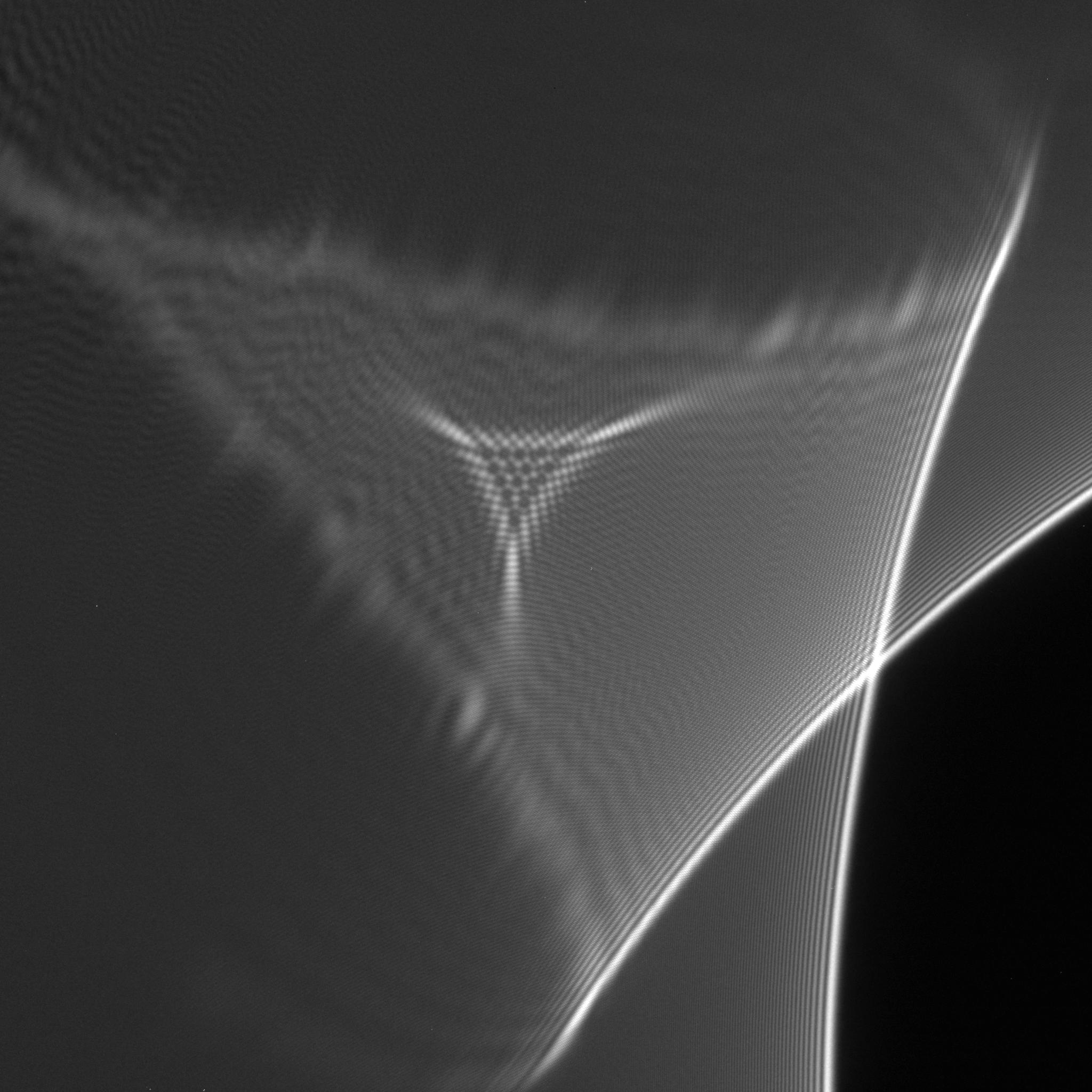
Fig. 1: Figure 1. Bright-field TEM image recorded at a defocus of 9.5 mm from two metallic needles that have a potential difference of 130 V between them. |
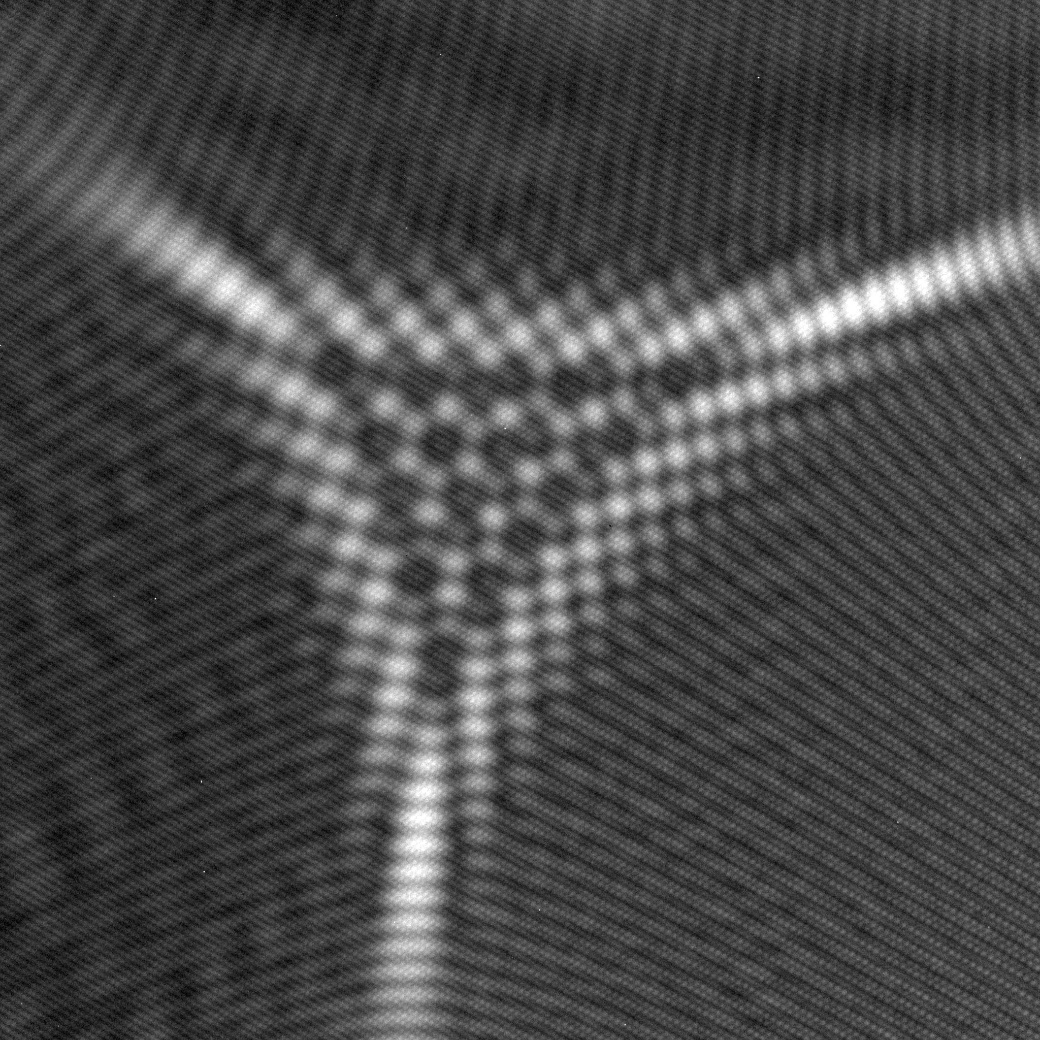
Fig. 2: Figure 2. Central region of Fig. 1 displayed at a higher magnification, revealing spots that have a hexagonal-like structure. |
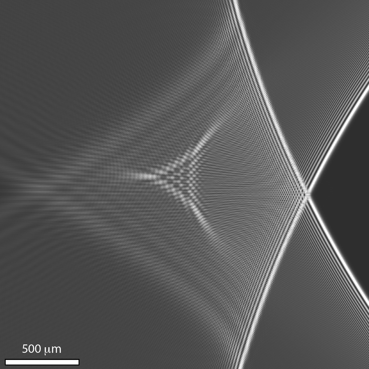
Fig. 3: Figure 3. Simulated image corresponding to the experimental conditions used to acquire Fig. 1. |
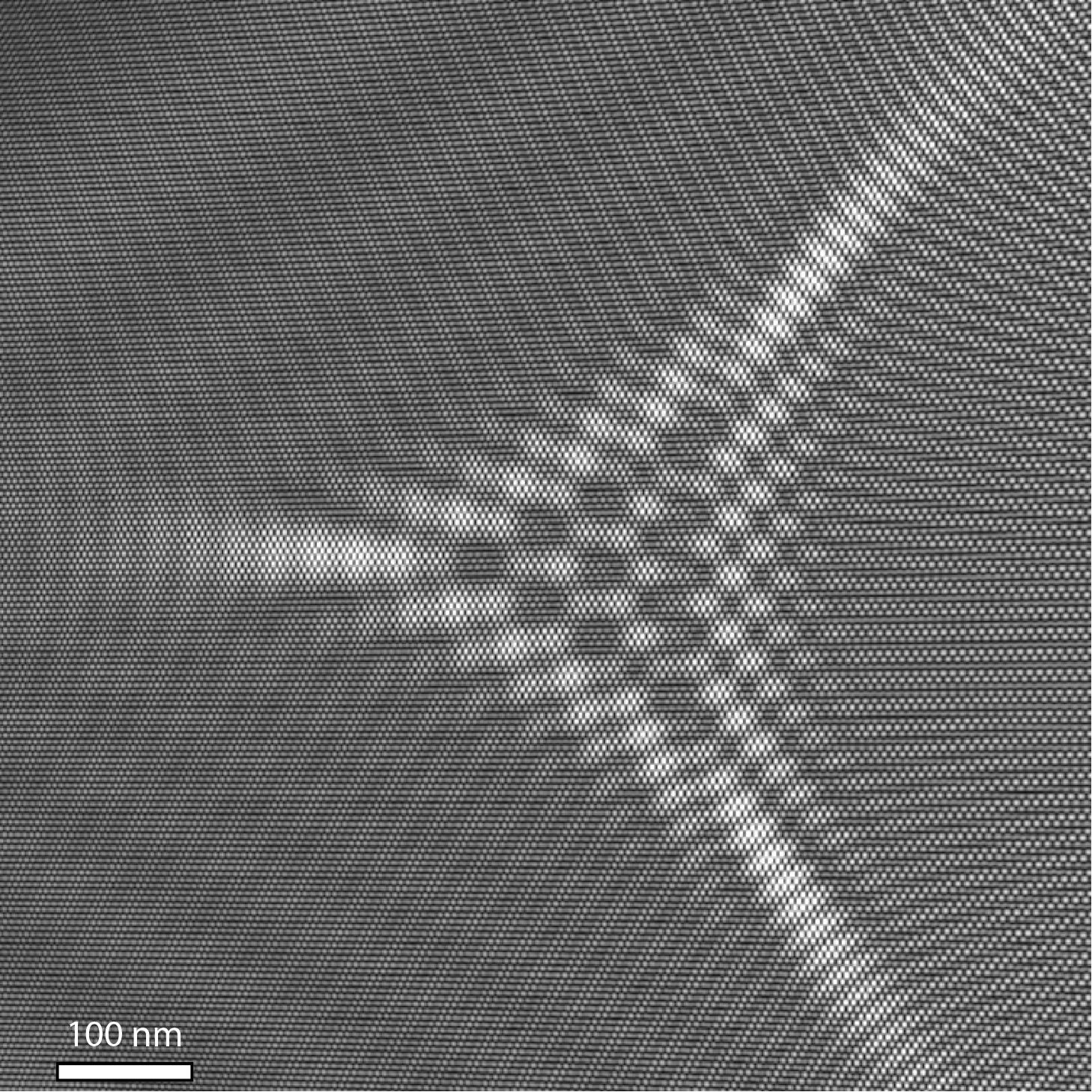
Fig. 4: Figure 4. Central region of Fig. 3 displayed at a higher magnification. |