IT-14-P-2566 DESIGN, IMPLEMENTATION AND CHARACTERIZATION OF A 3D-PRINTED AFM HEAD WITH PIEZOTUBE AND ELECTROMAGNETIC ACTUATORS FOR BIOMOLECULAR APPLICATIONS
An atomic force microscope (AFM) has been developed for biomolecular force spectroscopy. Design, implementation and characterization of the AFM head are described here. The head is portable and was manufactured at low cost using stereolithography. A flexible software-based controller was implemented that can be adapted easily for different applications. The AFM head, made of a rigid polymer material (Rigid Opaque, Stratasys, Ltd., MN, USA) is shown in Fig. 1(a). The head houses a piezotube actuator, a laser diode, a quadrature photodetector and an AFM cantilever. The cantilever is mounted to the piezotube using a customized holder. The incident laser beam (fiber pigtailed, Oz Optics, Ottowa, Canada) is directed to the cantilever using a kinematic mount. Reflected laser light is directed to the quadrature detector (Pacific Silicon Sensor, Westlake Village, CA, USA) via a mirror, which has a one degree-of-freedom of rotation. The photodetector is placed on a translational microstage with two degrees-of-freedom. The space below the cantilever plane is empty so that the head can be integrated with an inverted microscope. In addition, an electromagnet was employed with the head for direct cantilever actuation and for other magnetic applications. A one-dimensional actuation coil is integrated to the head as shown in Fig. 1(b). The system allows cantilever actuation using the piezotube actuator and the electromagnet. Fig. 2(a) shows a sample force curve obtained using the piezo actuator. The drive signal in various waveforms was generated by the customized software-based controller to actuate a commonly used AFM cantilever (SNL-10D, Bruker Probes, Santa Barbara, CA) on a silicon sample at various frequencies, from 10 mHz to 1 kHz. In addition, Fig. 2(b) shows a typical current signal input to the electromagnet the corresponding photodetector signal. A commercially available MFM cantilever (MESP, Bruker Probes, Santa Barbara, CA) was actuated by a square wave in air at 1 kHz. The head was designed and optimized for force spectroscopy experiments. The force noise of the system using typical AFM cantilevers has been characterized as 6.8 pN within a bandwidth of 1 kHz. Finally, a biomolecular force spectroscopy experiment to probe interactions between FGF-2 and heparin was performed using the piezotube actuation. Fig. 3(a) shows a typical force curve, exhibiting an unbinding event with a force strength of 500 pN, whereas in Fig. 3(b) there is another force curve indicating no adhesion/rupture events.
Authors would like to acknowledge funding from the EC (ICT FET-Open) under the MANAQA Project.
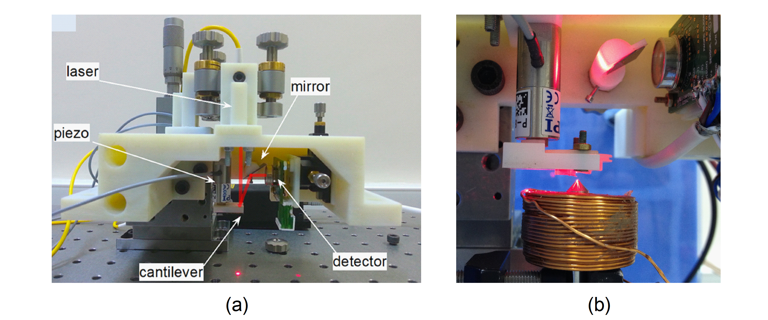
Fig. 1: Fig. 1 (a) The photograph of assembled AFM Head, manufactured using stereolitography. (b) The photograph of AFM head employed with an electromagnet to actuate the MFM cantilevers. |
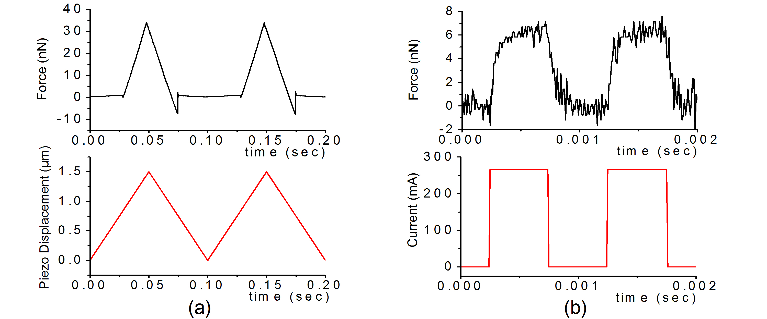
Fig. 2: Fig. 2 (a) Typical force curves, taken with 10 Hz piezo actuation over a silicon wafer. (b) Electromagnetic actuation of a MFM cantilever in air at 1 kHz. |
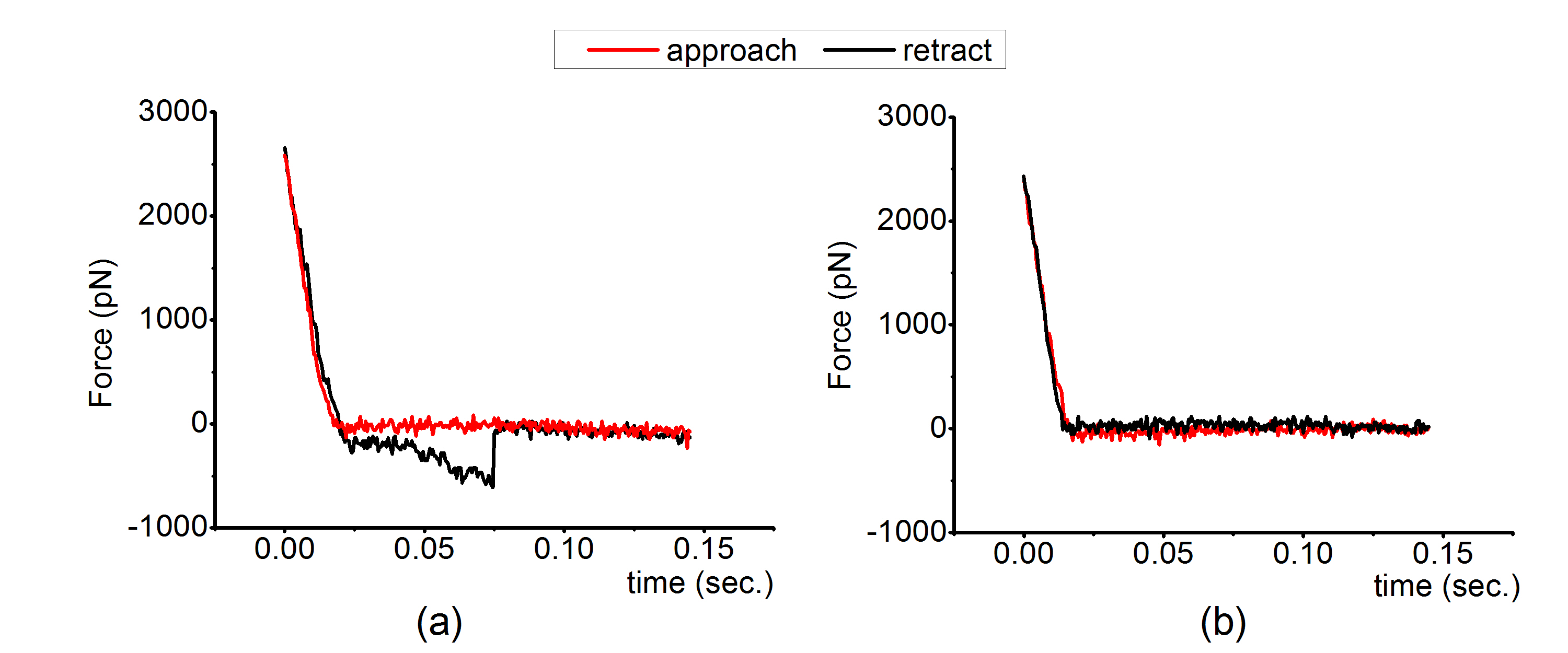
Fig. 3: Fig. 3 (a) A typical force curve exhibiting an unbinding event between FGF-2 and Heparin molecules. (b) A force curve which is indicating no adhesion/rupture event. |
|