LS-8-P-2514 RELIEF, SCHLIEREN AND PHASE-CONTRAST MICROSCOPY OF PLANTS: Label-free imaging and refractive index matching
INTRODUCTION
Detailed investigation of microscopic anatomy and its physiological implications often requires the use of specialized staining procedures and complex imaging equipment. In some cases, however, staining is impractical or even impossible, and contrasting by purely optical means represents a convenient alternative. The present paper highlights the advantages and pitfalls of several microscopic optical contrasting modalities suitable for imaging unstained plant cells/tissues and specimens derived from them.
APPARATUS
Relief- and schlieren-contrast imaging highlighted in the present paper utilize a shifting asymmetric (edge) diaphragm in the condenser and as such represent the simplest and historically also the oldest microscopic optical-contrasting modalities [1,2]. They are not directly available in most commercially available microscopes (see [3] for a rare exception), and one possible adaptation is depicted in Fig. 1. The rim of a partly pulled out objective Wollaston prism holder (itself a DIC-Nomarski imaging accessory) served as the schlieren diaphragm (simple modulator) at the objective back focal (Fourier) plane (Fig. 2).
RESULTS & DISCUSSION
Optically thick objects such as leaf replicas in transparent acrylate resin are typically best rendered in relief or schlieren contrast (Fig. 2), often superior in image quality to Hoffman modulation contrast, itself a more complex variant (requiring special objectives) of schlieren imaging. Other examples of relief-contrast (off-axis illumination) microscopy may be found elsewhere (e.g., [4]). Objects of medium optical thickness such as spores of the field horsetail (Equisetum arvense) or osmotically swollen (burst-open) pollen grains of yew or juniper are most conveniently imaged by Hoffman modulation contrast or apodized phase contrast. However, schlieren imaging yields superior contrast (especially when low-power objectives are used), comparable even to DIC-Nomarski microscopy (Fig. 3). Optically thin objects such as refractive-index matched stellar trichomes of olive (Olea europaea) or Elaeagnus sp. leaves ideally require the use of low-transmittance phase-contrast microscopy. Refractive-index matching (achieved by embedding, e.g., in acrylate resin or glycerol) is often essential to reliably visualize fine microscopic structures normally filled with air, such as the spokes of the seed-bearing ‘parachutes’ in the common dandelion (Taraxacum officinale), and makes it possible to determine their refractive index.
[1] Töpler A. (1866) Annalen der Physik 203 (4): 556-580
[2] Axelrod D. (1981) Cell Biophys. 3 (2): 167-173
[3] Hostounský Z. & Pelc R. (2007) Adv. Physiol. Educ. 31 (2): 232-235
[4] Pelc R., Hostounský Z. & Otaki T. (2008) J. Biomed. Opt. 13 (5): 054067
RP was supported by LC06063 and RVO:67985823 grants, and international NRF/KOSEF fellowship 2010. Some images were acquired on a microscope lent by Nikon CZ.
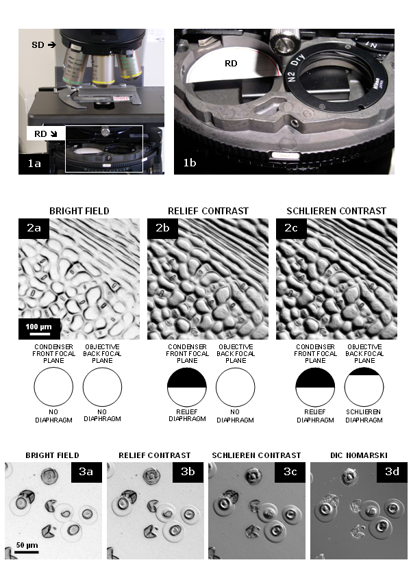
Fig. 1: Adaptation of Nikon D-CUD condenser to relief/schlieren imaging. Relief diaphragm (RD) made of black paper is used for imaging. SD, schlieren diaphragm. Fig. 2. Abaxial leaf replica of rhododendron (Daphniphyllum macropodum) in transparent resin (image ID: 2010-02-24_*). Fig. 3. Pollen grains of yew (Taxus baccata) in water (image ID: 2009-08-29_*) |