IT-2-P-2453 3D-structural elucidation of highly ordered mesoporous TiO2 thin film by the method of electron crystallography
At present, transmission electron microscopy 3D reconstruction has become an important technique to elucidate the 3D structure of materials. Electron tomography, single particle analysis and electron crystallography are the common methods of 3D reconstruction. Thereinto, electron crystallography, comprising transmission electron microscopy and electron diffraction, determines the 3D structure of materials via their 2D structure information. This method is used to investigate the structure of crystal in general. In fact, due to the similar structure between highly ordered and crystal, highly ordered mesoporous structure can also form electron diffraction patterns, so the method of electron crystallography can be used to study the 3D structure of highly ordered mesoporous TiO2 thin film as well. In this experiment, we obtained TEM images and their corresponding slected-area electron diffraction (SAED) patterns from different zone axes. Especially, the corresponding SAED patterns were recorded at an instrument camera length of 200 cm, which is much longer than the average length. The top view TEM image of the film shows a highly ordered honeycomb arrangement with a nearly perfect hexagonal disposition. Its corresponding SAED pattern exhibits a 6-fold symmetry, which is compatible with the [001] zone axis of the hexagonal structure. Mainly, the diffraction patterns taken from three directions ([001], [1-10] and [121]) can be indexed in the P63/mmc space group. And the cross-section TEM image regarded as viewed from [100] zone axes shows an ABAB stacking sequence. Moreover, the diameter of the pores can be directly measured to be ~10nm. In summary, the method of electron crystallography implements an effective explanation of highly ordered mesoporous TiO2 thin film with 3D hexagonal structure.
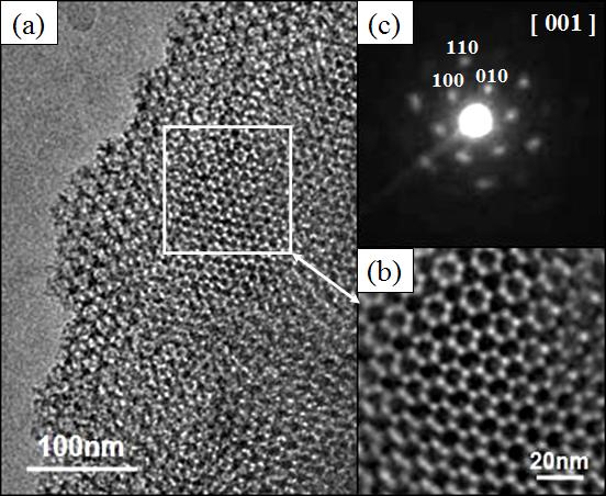
Fig. 1: The TEM images along [001] with SAED |
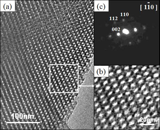
Fig. 2: The TEM images along [1-10] with SAED |
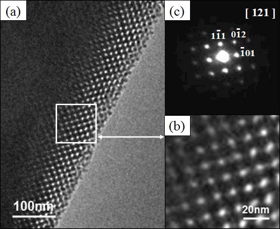
Fig. 3: The TEM images along [121] with SAED |
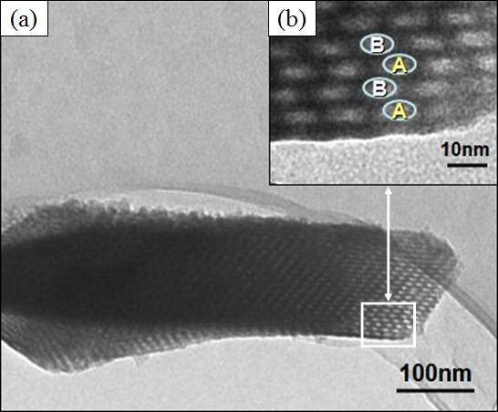
Fig. 4: The cross-section TEM images along [100] |