IT-6-P-2451 Aberration-Corrected, Environmental TEM Studies on Carbon Nanotube Oxidation and the Influence of the Imaging Electron Beam
One of the major applications for carbon nanotubes (CNTs) is as field emission electron sources, for example in image displays and high-intensity medical X-ray tubes [1-3]. The emission currents and lifetimes of CNTs are found to decrease under less stringent vacuum conditions [4, 5]. Earlier reports of carbon nanotube oxidation performed in an external laboratory setting, and surveyed a posteriori with a transmission electron microscope (TEM), suggested that the nanotube caps were selectively attacked during the oxidation process [6, 7].
Recently, we reported the direct study on the structural changes in CNTs as they were oxidized in-situ using an aberration-corrected environmental TEM (ETEM) [8]. Nanotubes were identified and tracked for structural changes as they were heated to increasing temperatures in a 1.5 mbar, high purity (99.9999%) oxygen environment. In order to investigate the effect of gaseous oxygen molecules on the nanotubes, rather than ionized gas species, we established a protocol whereby heating and oxidation were performed without an imaging beam, and the changes on identifiable nanotubes were documented after purging the gas from the chamber. Our studies showed that the oxidation of multiwall CNTs proceeds layer by layer, starting with the outermost wall, and not initiating at the nanotube cap, as reported previously. Nanotubes with a larger number of walls (greater than six) were found to be more resistant to oxidation, with all walls remaining intact during the ETEM experiments [8].
To simulate the highly ionized environment, which is expected during field emission, we repeated these observations at room temperature in the ETEM in the presence of the imaging beam. Under such conditions, we found that more rapid attack takes place, even at room temperature, and both the hemispherical cap and side walls of the CNTs are vulnerable. The influence of the imaging electron beam in the observation of this gas-solid reaction in the ETEM will be discussed.
References:
[1] Q. H. Wang et al., Appl. Phys. Lett. 72 (1998) pp. 2912–2913
[2] G. Cao et al., Med. Phys. 37 (2010), pp. 5306–5312
[3] X. Qian et al., Med. Phys. 39 (2012), pp. 2090–2099
[4] K. A. Dean and B. R. Chalamala, Appl. Phys. Lett. 75 (1999), pp. 3017–3019
[5] J.-M. Bonard, et al., Ultramicroscopy 73 (1998), pp. 7–15
[6] P. M. Ajayan et al., Nature 362 (1993), pp. 522–525
[7] S. C. Tsang, P. J. F. Harris and M. L. H. Green, Nature 362 (1993), pp. 520–522
[8] A. L. Koh et al., ACS Nano 7(3) (2013), pp. 2566–2572
Funding from the National Cancer Institute grants CCNE U54CA-119343 (O.Z.), R01CA134598 (O.Z.), and CCNE-T U54CA151459-02 (R.S.) is acknowledged. We thank Dr. Bo Gao of Xintek for providing the CNTs.
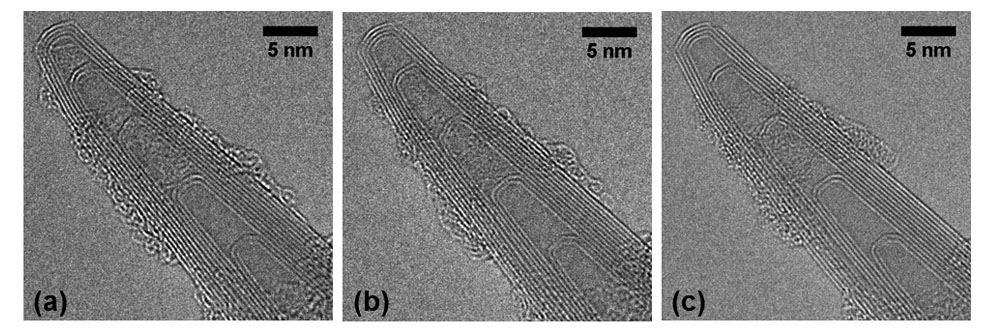
Fig. 1: Aberration-corrected TEM images of multiwall carbon nanotubes (MWNT) at (a) 400°C, (b) 400°C after exposure to 1.5 mbar oxygen for 15 min, and (c) 520°C after exposure to 1.5 mbar oxygen for 15 min. The electron beam is blanked during the oxidation process. The MWNT is found to resistant to oxidation under these conditions. |
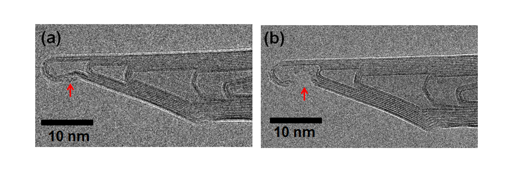
Fig. 2: Aberration-corrected TEM images of multiwall carbon nanotubes (MWNT) acquired at room temperature and 1.0 mbar oxygen, with the imaging electron beam on. The red arrow indicates attack on the MWNT cap and side wall, due to exposure to the ionized oxygen. The time elapsed between (a) and (b) is 32 sec. |