IT-10-P-2426 Nonlinear intensity attenuation in bright-field TEM images and its influence on tomographic reconstruction of micron-sized materials
Currently, tomography in transmission electron microscopes (TEM) is widely applied to three-dimensional (3D) analyses of nanometer-sized and sub-micron-sized materials. One of the next methodological targets should be quantitative 3D reconstructions in which not only the shape but also the internal density are correctly reproduced. This is hindered generally by the nonlinearity between projection thickness and image intensity. In the case of mass-thickness contrast in bright-field TEM (BF-TEM) images, the ideal exponential attenuation of the image intensity with increasing thickness is disturbed by multiple scatterings.
In the present study, the nonlinear attenuation in BF-TEM images was analyzed using amorphous carbon microcoils (CMCs) [1] shown in Fig. 1(a). Their well-defined shapes and compositional homogeneity are quite useful for estimating the mass-thickness [2]. The intensity attenuation was measured along the line in Fig. 1(b), which was taken by the high-voltage electron microscope in Nagoya University [3]. The results measured at the acceleration voltages of 400, 600, 800 and 1000 kV were converted to the plots of the electron transmittance T in Fig. 2. At a glance, T at any voltages seems to undergo the linear attenuation. However, the least squares fitted line for the data at 400 kV exhibits a considerably negative intercept value at zero thickness. Such nonlinear attenuation should induce failures in conversion from intensity to thickness and thus inhibits correct 3D reconstructions of the internal density.
The influence of the nonlinearity on tomographic reconstructions was examined using a 360°-tilt sample holder, which was specially developed for eliminating the missing-wedge effect [2]. Figure 3 shows the results of the reconstructions from the tilt series taken at 400 kV and 1000 kV. Although the 3D shape of the CMC has been reconstructed well in both cases, the internal density is not uniform but has a gradient from the center at 400 kV. Moreover, there is a slight increase in the vacuum level in the interior of the coil. The inaccurate density reconstruction should result from the nonlinearity shown in Fig. 2. Judging from the plot for 600 kV electrons in Fig. 2, the linearity is valid at least down to lnT = −0.4, which corresponds to the electron transmittance of about 2/3. This information should be beneficial in practical tomography experiments because one can foresee quality of the reconstruction from the minimum transmittance in a single BF-TEM image prior to the tilt series acquisition.
References
[1] S. Motojima et al., Appl. Phys. Lett. 56 (1990) 321.
[2] J. Yamasaki et al., submitted to Microscopy.
[3] N. Tanaka et al., Microscopy 62 (2013) 205.
The authors are grateful to Mr. M. Ohsaki in JEOL Ltd. for discussions about designing the sample holder and System In Frontier Inc. for discussions on precise 3D reconstruction procedures. We also thank Mr. Y. Yamamoto and Dr. C. Morita of HVEM laboratory in Nagoya University for their assistance with the experiments. One of the authors (N.T.) thanks Dr. S. Motojima for useful discussions.
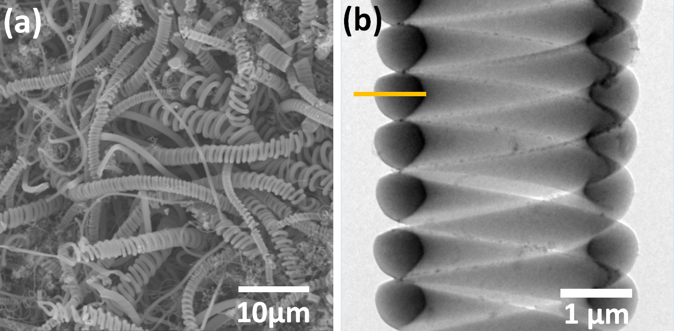
Fig. 1: Carbon microcoils. (a) SEM image (Microphase Co., Ltd.) and (b) BF-TEM image taken by the HVEM. |
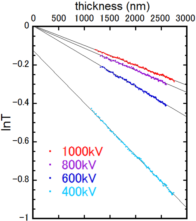
Fig. 2: Attenuation of electron transmittance T in BF-TEM images with increasing thickness. |
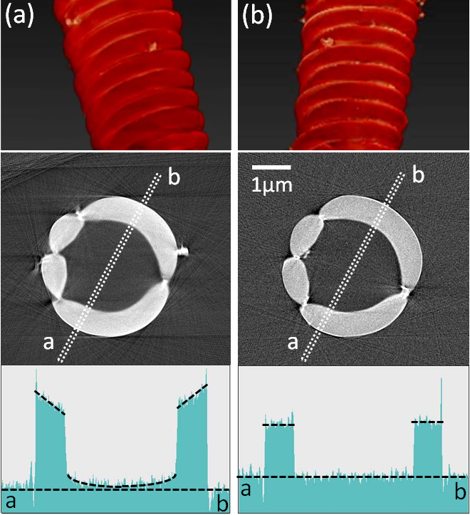
Fig. 3: 3D reconstructions of the CMC from the tilt series of BF-TEM images taken at (a) 400 kV and (b) 1000 kV. |
|