IT-11-O-2391 Electron exit wave reconstruction in Gaussian basis: from high resolution image to diffraction pattern
Accurate electron exit wave reconstruction can offer not only increased resolution and improved signal to noise ratio but it can also provide some 3D information about the sample in high resolution transmission electron microscopy (HRTEM). Restoration of the exit wave from experimental data usually involves collecting a series of images with varying focus (a focal series). The exit wave can be also recovered by recording overlapping diffraction patterns (DP) and applying specially designed iterative numeric algorithms in a ptychographic approach. Both these approaches require a number of images or diffraction patterns to be collected, which increases the total electron dose that the sample needs to withstand. Such an approach can be difficult to implement for radiation sensitive materials, due to possible sample damage during acquisition of focal or diffraction series.
In the present work we suggest and test an approach that in principle allows reconstructing exit wave from a single image or diffraction pattern for a weak-phase object. One of the obstacles to using only one image or diffraction pattern for the reconstruction is that in this case the number of variables in the exit wave to be determined is comparable to the number of data recorded. Presence of the aberrations of the objective lens and experimental noise further complicates the analysis and can lead to multiple or unstable solutions. By representing the electron exit wave in Gaussian basis we greatly reduce the number of variables needed to find the solution. This approach also has an advantage of analytic representation of the DP and also derivatives needed for the reconstruction process. We test the suggested method on experimental HRTEM image of graphene and simulated diffraction pattern of a carbon nanotube. The reconstruction algorithm involves solving an overdetermined system of non-linear equations with either numeric or analytic derivatives and appears robust to noise. We compare the reconstructed experimental exit wave phase from graphene with the multislice simulations.
We thank the financial support from the European Union under the Seventh Framework Program under a contract for an Integrated Infrastructure Initiative (Ref 312483-ESTEEM2).
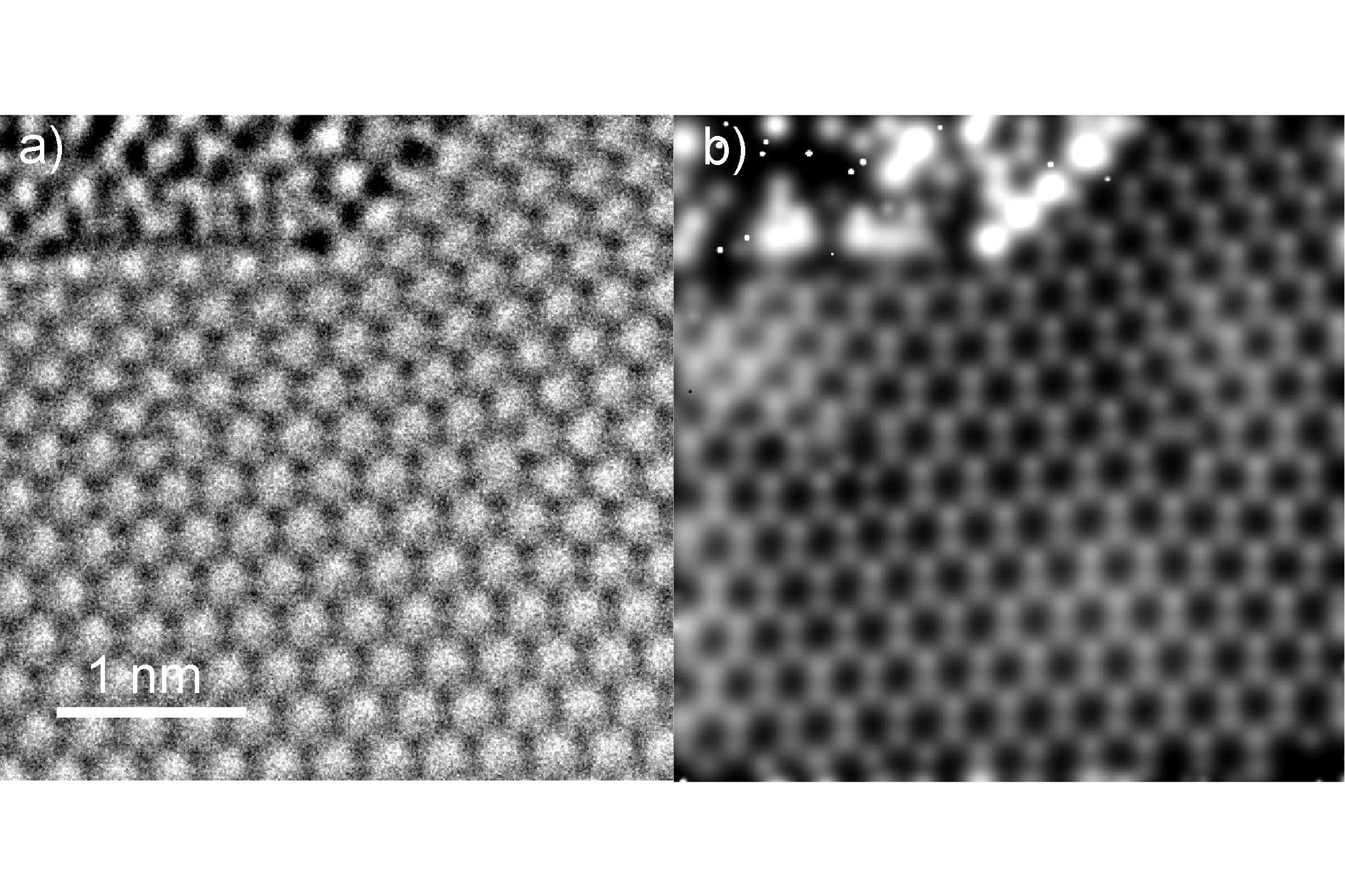
Fig. 1: Experimental image of graphene a) and the reconstructed exit wave phase b). |
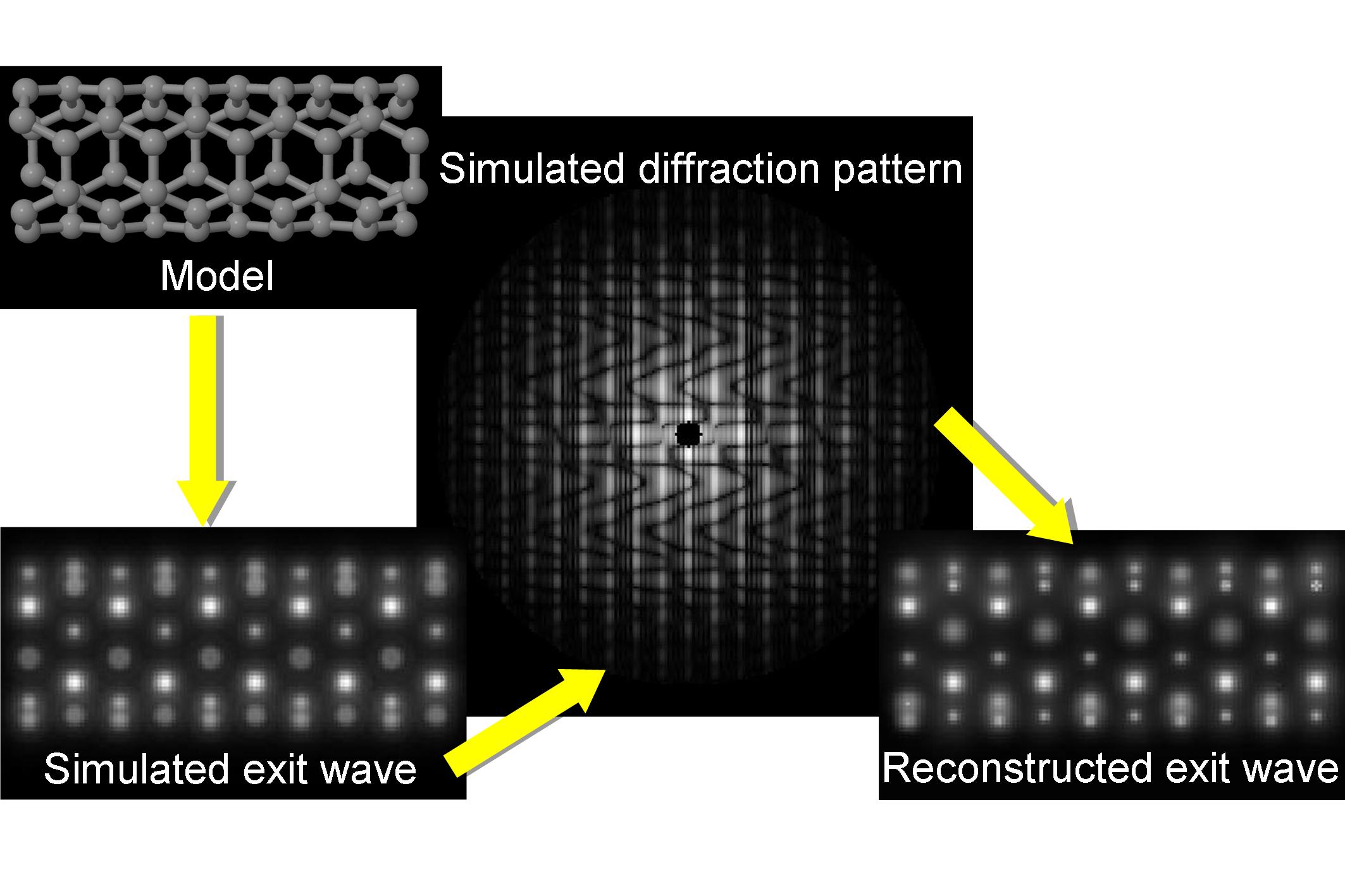
Fig. 2: Simulated input exit wave and corresponding target diffraction pattern (represented as a logarithm of the square root of the diffracted intensity) and the reconstructed exit wave. |