IT-10-P-2280 Tomography in Analytical Transmission Electron Microscopy of Nanomaterials
The engineering of specific material properties requires both a structural and chemical understanding, often obtained with electron microscopic techniques. While analytical scanning transmission electron microscopy (STEM), including energy dispersive x-ray (EDX) spectroscopy and electron energy loss spectroscopy (EELS), can offer the necessary chemical information, the integrative character of the signal acquired through transmission might hide important structural details of the material. Those details can be revealed by using electron tomography, where the data is acquired at different tilt angles and, after alignment, reconstructed to form a full 3D model of the investigated material. This technique on its own, however, lacks the important chemical information. The content of this work is the combination of both techniques, analytical STEM and tomography, which offers a more complete understanding of the material structure and composition.
Combining analytical signals in the form of spectrum images with a tomographic acquisition however, represents a major challenge. First of all there is no possibility to acquire a tilt series including EELS and EDX spectra for each data point automatically. Secondly, there is no simple way of reconstructing a four dimensional object, containing two spatial, one energy and one tilt angle coordinate. Having tackled this problem, the next issue arises due to the often very limited statistical quality of the analytical signals. In order to minimize acquisition times, dose and sample drift, dwell times per pixel are often in the order of a few milliseconds, necessitating special reconstruction algorithms that can handle noisy data.
The material studied in this work is an alloy containing scandium and zirconium rich nanoparticles embedded in an aluminum-magnesium matrix (fig.1 and fig.2). These nanoparticles increase the mechanical resistance of the material. Their sizes and chemical compositions can vary, depending on the aging process. Previous work reported that these particles can exhibit a core-shell structure.
As the acquisition of EDX and EELS spectra takes more time than non-spectroscopic imaging techniques special care needs to be taken concerning the stability of the sample. This can be achieved by reducing the number of tilt angles, which is possible if special reconstruction algorithms are used. Total variation minimization mathematically assumes minimized gradients which can reduce artefacts and noise. While this can be problematic if the sample itself exhibits gradients of concentrations, it can lead to smooth reconstructions if the sample consists of clearly separable phases.
We thank the Austrian Cooperative Research Facility, the European Union (7th Framework Programme: ESTEEM2), and the Austrian Research Promotion Agency FFG (TAKE OFF project 839002) for funding.
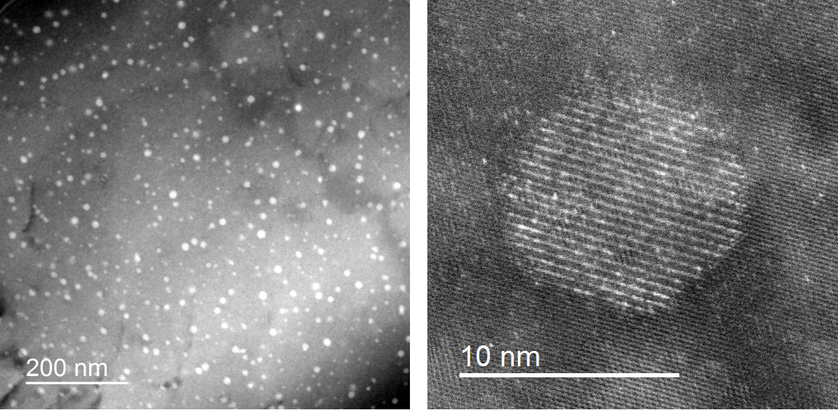
Fig. 1: aluminum alloy; left: EFTEM overview image at 40eV; right: HAADF image of Sc-rich nanoparticle |
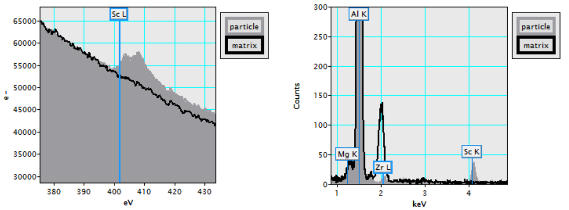
Fig. 2: extracted spectra of an EELS (left) and EDX (right) spectrum image of a nanoparticle in its matrix, supporting that the particle is Sc-rich |