IT-2-O-2221 Channeling Effects on the Accuracy of HAADF STEM Quantification of Bimetallic Catalyst Nanoparticles
Quantification of high angle annular dark-field scanning transmission electron microscope (HAADF STEM) images uses atomic resolution images as data sets to extract sample composition and thickness information from. A new quantification method based on calculating the scattering cross-section (CS) of each atomic column has been shown to be more robust to microscope image parameters.1 Using an automated code,2 this analysis involves converting images to an absolute scale through detector normalisation3, integrating over each atomic column within an image and multiplying by pixel area.
Whilst mathematically robust to microscope imaging parameters there are many other factors which affect the accuracy of quantification results. Channelling occurs when the columns of atoms in a crystal are aligned parallel to the incident electron beam and they act like miniature lenses providing an extra focusing effect on the probe. The subsequent atoms in the atomic column see a more focused probe than the first atom; resulting in them supplying increased scattering out to the detector. Along the length of the column, oscillations in intensity are seen, much as though the electrons are propagating in a waveguide. The whole column may therefore have a different scattering CS than the sum of the individual CSs of its constituent atoms. The ordering of atom types within an atomic column also affects the overall CS. Comparably another process known as de-channelling provides cross-talk between neighbouring columns of atoms. Cross-talk occurs when part of the probe is scattered and becomes channelled by a neighbouring column of atoms and then scattered out to the detector, thereby contributing information to the signal from neighbouring columns.
Atomic resolution requires viewing a crystal down a low order zone axis; any sample mis-tilt away results in a reduction in the channelling contribution and therefore a loss in CS, Figure 1. By 4̊ of tilt the effects of channeling are almost completely lost, whilst some atomic resolution remains. Top-bottom effects in the bimetallic columns are also diminished by 4̊ mis-tilt. At small tilts, however, there is a plateau region where the CS is independent of tilt, the size of which is dependent on probe convergence angle size, Figure 1. We believe the robustness to tilt when imaging on axis is more beneficial than the potential composition information gain from tilting far off a zone axis. This is particularly the case for nanoparticles which tilt under the beam. Combining with spectroscopy techniques will be necessary for gaining compositional information.
1 H E et al, Ultramicroscopy 133 (2013), p109-19
2 The Absolute Integrator is free for academic use from www.lewyjones.com/software/
3 JM LeBeau et al, Nano Letters 10 (2010), p4405-8
The research leading to these results has received funding from the European Union Seventh Framework Programme under Grant Agreement 312483 - ESTEEM2 (Integrated Infrastructure Initiative–I3), and from the EPSRC (grant number EP/K032518/1).
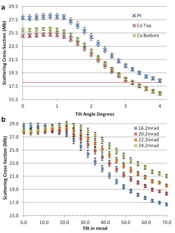
Fig. 1: The CS of a column of 7 Pt atoms in a crystal plotted against tilt away from a <110> zone axis. (a) Pt, blue, with the top or bottom atom replaced with Co, red and green, show a plateau region before dropping with tilt. With no channelling the CS would be 7x1 Pt atom, dotted red. (b) Tilt plot with different probe convergence angles. |