IT-3-P-2211 Flexible Structured Illumination Microscope with a Programmable Illumination Array
Structured illumination microscopy (SIM) has grown into a family of methods which achieve optical sectioning (OS-SIM), resolution beyond the Abbe limit (SR-SIM), or a combination of both effects in optical microscopy. SIM techniques rely on illumination of a sample with patterns of light which must be shifted and/or rotated between each acquired image. The patterns are typically created with physical gratings or masks, or by laser interference, and the final optically sectioned or high resolution image is obtained computationally after data acquisition. We used a flexible, high speed ferroelectric liquid crystal display for definition of the illumination pattern coupled with widefield detection and subsequent image processing. Focusing on optical sectioning, we developed a unique and highly accurate calibration approach which allowed us to determine a mathematical model describing the mapping of the illumination pattern from the microdisplay pixels to the camera sensor pixels. This is important for higher performance image processing methods such as scaled subtraction of the out of focus light, which require knowledge of the illumination pattern position in the acquired data. The calibration is also advantageous for SR-SIM reconstruction, as it provides precise information about reconstruction parameters (pattern period, orientation and phase) [1]. We evaluated the signal to noise ratio and the sectioning ability (see Fig. 1) of the OS-SIM reconstructed images for several data processing methods and illumination patterns with a wide range of spatial frequencies [2].
[1] M. G. L. Gustafsson, “Surpassing the lateral resolution limit by a factor of two using structured illumination microscopy,” J. Microsc., vol. 198, pp. 82–87, 2000.
[2] P. Křížek, I. Raška, and G. M. Hagen, “Flexible structured illumination microscope with a programmable illumination array.,” Opt. Express, vol. 20, no. 22, pp. 24585–99, Oct. 2012.
This work was supported by the Grant Agency of the Czech Republic [P304/09/1047, P205/12/P392, P302/12/G157 and 14-15272P], by Charles University in Prague [Prvouk/1LF/1, UNCE 204022], and by European Union Funds for Regional Development [OPPK CZ.2.16/3.1.00/24010].
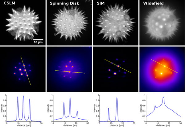
Fig. 1: Comparison of different optically sectioning microscopes on a pollen grain sample. The SIM system achieves an optical sectioning thickness of 300 nm, much better than is possible in CLSM (Confocal Laser Scanning Microscopy). |