IT-5-P-2163 An ELNES study of anisotropic materials using variable beam energies
The development of (S)TEMs in recent years is pointing towards a higher variability of beam energies. The reasons are manifold, as there are amongst others less beam damage [1], larger elastic and inelastic scattering cross sections, and less or even no excitation of Cerenkov losses for the analysis of optical properties [2,3]. Simultaneously the development of electron detectors being able to handle slower electrons efficiently allows detecting low signals with little noise. This is important, because low voltage electron beams have usually little beam current.
In the present work we demonstrate the effect of the varying beam energy on the ELNES of the B-K edge caused by the change in momentum transfer with respect to the forward (qz) and perpendicular (qperp) directions. With decreasing beam energy qperp is stronger decreasing than qz. For the experiment we orient the c-axes of a hexagonal BN crystal parallel to the electron beam. For low beam energies the van der Waals bonds contribute to the EELS spectrum stronger as compared to high beam energies showing a small qz. This can be seen in Figure 1, because the π* peak decreases with increasing beam energy. When orienting the beam axes perpendicular to the c-axes of h-BN the effect is inverted and the sp2 orbitals are contributing stronger with decreasing beam energy (see Figure 2).
We compare the experiments with ab initio calculations using the Wien2k code. Using the TELNES.3 routine, the effects of orientation dependence [4] as well as the influence of the beam energies on the fine structure of the Boron K-edge can be simulated. One can investigate orbital dependent properties by comparing these simulations to the experimentally acquired spectra. As changing the beam energy is in some cases a much easier task to perform than conducting ELCE experiments [5], this technique can be an alternative or a complementary procedure.
[1] U. Kaiser et al., Ultramicroscopy 111 (2011), 1239 - 1246
[2] M. Stöger-Pollach, Micron 39 (2008), 1092 - 1110
[3] M. Stöger-Pollach, Micron 41 (2010), 577 – 584
[4] C. Hebert-Souche et al., Ultramicroscopy 83 (2000), 9 – 16
[5] W. Hetaba et al., Micron, in press.
The authors aknowledge the USTEM facility for providing the low-KV TEM.
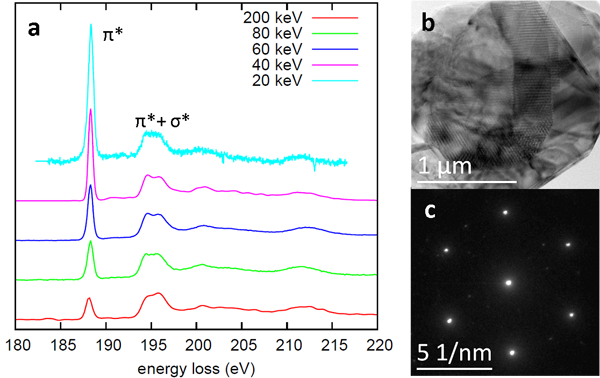
Fig. 1: Figure 1: a) Boron-K edge of BN recorded with various incident beam energies (normalized to the 195.8 eV peak). b) Bright field image of the BN specimen. c) Diffraction pattern of h-BN showing the [0001]-orientation. Consequently the c-axes of the h-BN is parallel to the beam axes. |
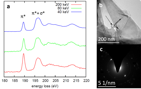
Fig. 2: Figure 2: a) Boron-K edge of BN recorded with various incident beam energies (normalized to the 195.8 eV peak). b) Bright field image of the BN specimen. The circle indicates the position of the EELS experiments. c) Diffraction pattern of h-BN showing the (0002) spots only. Consequently the c-axes of the h-BN is perpendicular to the beam axes. |