IT-8-P-2148 Sub-picosencond electron beam and femtosecond optical pump system in spin-polarized TEM
Time-resolved measurements at nanometer spatial resolutions are very important for investigating relaxation processes, catalyzed reactions and phase-transition phenomena. It is possible to carry out such time-resolved analysis using transmission electron microscopy (TEM) by using a pulsed electron beam as a probe beam. Such an approach has been applied in dynamic TEM (DTEM) and ultra-fast electron microscopy (UEM), which use metals and LaB6 for a photoemission source driven by a pulsed laser. These methods have led to the possibility of four-dimensional electron microscopy with high spatial and temporal resolutions. Spin-polarized transmission electron microscopy (SP-TEM) can satisfy two abilities of spin-resolved imaging and pulsed electron gun operation simultaneously, because the instrument consists of a laser-driven polarized electron source (PES) and a conventional TEM system [1].
Spin-polarized electron can be generated by photoemission from III–V semiconductors with a negative electron affinity (NEA) surface. Several beam parameters of a PES are vastly superior to those for conventional thermal electron beams. A high spin-polarization of 92% and a high quantum efficiency of 0.5% have been simultaneously realized using a GaAs–GaAsP strained-layer-superlattice photocathode. In addition, such a photocathode has the ability to generate a sub-picosecond multibunch beam [2]. In order to realize a pump-probe method using the spin-polarized pulse electron beam, we have developed a synchronizing system and demonstrated a phase-locked TEM image of wobbling probe beam by using a pulse electron beam [3]. TEM images were already acquired with a pulsed electron beam with a 1.4-ns pulse duration. Now we have constructed a new optical system, which can provide a sub-picosecond pulse laser and femtosecond pulse simultaneously, to realize an ultrafast temporal resolution. The figure 1 (b) and (c) show the photograph and the schematic diagram, respectively. The sub-picosecond pulse laser is used to drive the electron gun. Another femtosecond laser is transferred to pump a specimen to create an excited state. The sub-picosecond pulse is generated by narrowing a bandwidth of a seed laser which is emitted from a mode-lock Ti:Sapphire laser. Figure 2 shows the femtosecond and picosecond pulse. The sub-picosecond pulse laser is necessary to keep the high polarization. These results suggest the possibility of pump-probe measurements in SP-TEM using the pulsed electron beam as a probe, allowing nanometer-scale time-resolved spin mapping.
[1] M. Kuwahara et al., Appl. Phys. Lett. 101 (2012) 03310
[2] Y. Honda, et al., Jpn. J. Appl. Phys. 52, 086401-086407(2013).
[3] M. Kuwahara et al., Microscopy 62, 607-614 (2013).
The authors thank Drs. H. Shinada, M. Koguchi and M. Tomita of Hitachi Central Research Laboratory for fruitful discussions and encouragement. This research was supported by MEXT KAKENHI Grant Number 51996964, 24651123, 25706031.
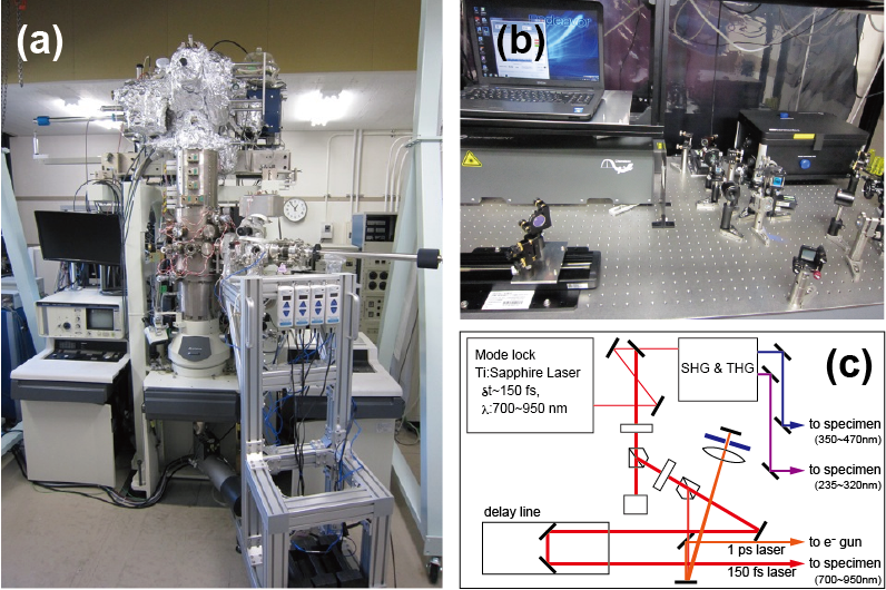
Fig. 1: (a) Photograph of the spin-polarized TEM, (b) Photograph of pulse laser system and (c) the schematics. |
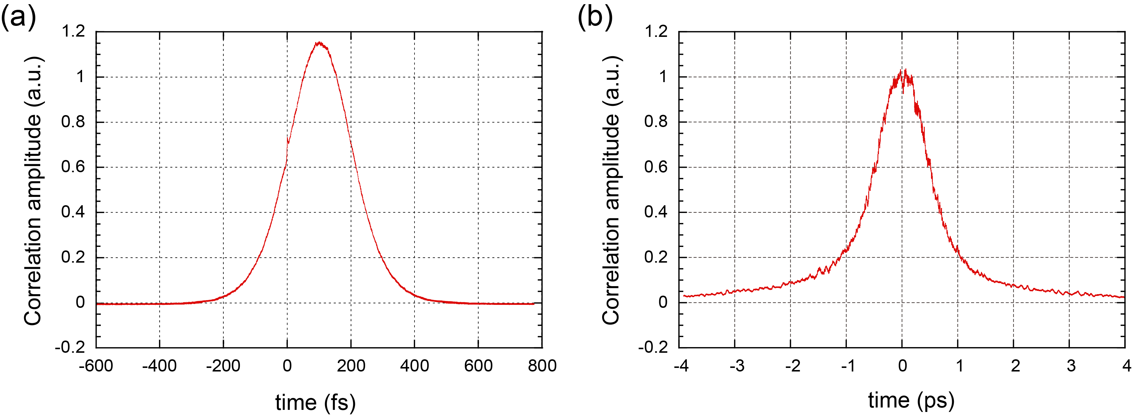
Fig. 2: Auto-correlation amplitudes of pulse lasers as a function of correlation time. (a) a correlation amplitude of a femtosecond pulse laser for excitation of a specimen. (b) a correlation amplitude of picosecond pulse laser for driving an electron source. |