IT-11-P-2111 Optimisation of spatial and phase resolution of off-axis electron holography for detection of single dopant atoms.
The reduction of the size of semiconductor devices leads to use of higher dopant concentrations. The scale of these devices implies that what was known as a very high dopant concentration will be only a few tens of atoms. The small numbers of these dopants mean that the just one of these atoms out of place will become statistically significant with respect to the electrical properties of the device. Thus, it is becoming increasingly important to be able to characterise the position and activity of these individual dopants.
Assuming a perfect specimen and vacuum, simulations show a phase sensitivity of better than 2π/2000 will be required in order to detect a single ionised dopant atom in silicon. The current state of the art for atomic resolution is around 2π/200, thus an improvement of an order of magnitude is required. The phase sensitivity of a reconstructed phase image is given by the relation,
Δφ≈(2/NV2)0.5
where N is the number of electron counts and V is hologram contrast. In order to achieve the best spatial resolution at atomic resolution it will not be possible to acquire holograms for very long periods due to specimen drift. Thus different approaches must be employed to improve the sensitivity.
Off-axis electron holography has been performed using a double aberration corrected FEI Titan Ultimate TEM equipped with an X-FEG. To improve the number of counts, a large series of holograms can be added together [1]. Fig. 1 shows a phase image of a silicon calibration specimen delta-doped with boron atoms. In Fig. 1(a) a single phase image acquired for 8 seconds is shown whereas (b) shows improvements from summing 25 holograms. In order to improve the contrast of the holograms, a monochromator can be used. Fig. 2 shows the effects of using the monochromator on the fringe contrast measured on reference holograms. An increase from 25 to 35 % will make a significant step towards the target of 2π/2000. Other approaches have been used to improve the reconstruction procedure. The spatial resolution of holograms containing strong phase objects is usually one third of the carrier frequency to avoid cross-talk from the centreband. The suppression of centreband in Fourier space by using phase shifting holography [2] improves the spatial resolution for a given carrier frequency and relaxes the electron biprism bias, resulting in higher V.
In this presentation we will show how combinations of these different methods of optimising phase noise and spatial resolution have been combined and then applied to wedge polished silicon specimens that contain different types of dopant atoms.
[1] R. McLeod et al. In press Ultramicroscopy (2013)
[2] V. Volkov et al. Ultramicroscopy 134 p175 (2013)
This work has been performed on the nanocharacterisation platform (PFNC) at Minatec. DC thanks the European Research Council for the Starting Grant “Holoview”.
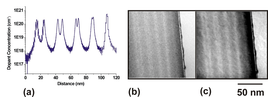
Fig. 1: (a) Secondary Ion Mass Spectrometry (SIMS) profile of boron doped delta layers. (b) Reconstructed phase image from single hologram acquired for 8 seconds. (c) Reconstructed phase image from 25 holograms summed together. |
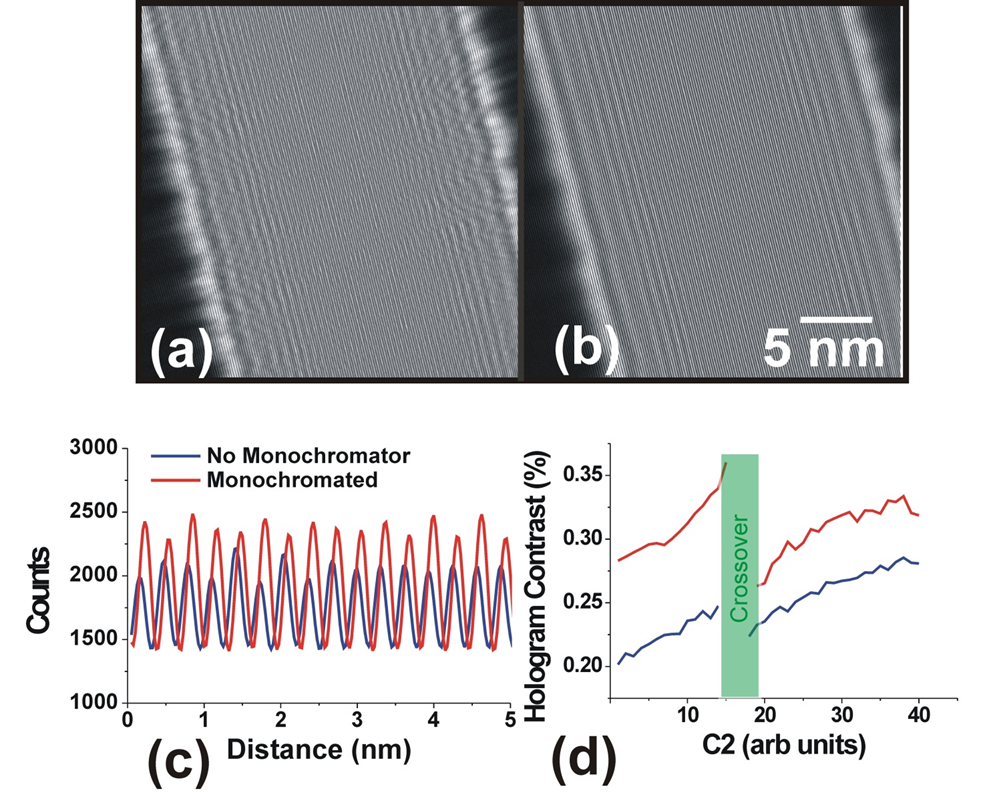
Fig. 2: Electron holograms acquired without (a) and with (b) monochromator active. (c) Fringe intensity profiles acquired from hologram and (d) measured contrast as a function of C2 with and without monochromator. |