IT-7-P-2081 In Situ 3D Studies of the Chlamydomonas Chloroplast Using Cryo-Focused Ion Beam Milling and Cryo-Electron Tomography
A comprehensive understanding of eukaryotic photosynthesis, the process that converts light energy into biochemical energy, requires a molecular-resolution three-dimensional model of the chloroplast’s intricate structure. Although the first transmission electron microscopy (TEM) studies of this important organelle date back to the early days of TEM in the 1950s, these observations, and the subsequent studies in the following decades, were limited by artifact-inducing sample preparation techniques. While valuable knowledge has been gained by both freeze-fracture and conventional heavy-metal stained plastic section preparations, the three-dimensional native architecture of the chloroplast can only be visualized by cryo-electron tomography (cryo-ET) of vitreous samples. In situ cryo-ET of specific subsystems within larger eukaryotic specimens requires selected areas of vitreous material to be thinned to electron transparency (less than 500 nm). Until recently, cryo-sectioning with an ultramicrotome was the only method capable of achieving this goal. However, cryo-ultramicrotomy is a laborious and technically demanding technique, and furthermore, mechanical sectioning introduces inevitable artefacts such as compression deformations.
In this work, we show that cryo-focused ion beam (cryo-FIB) milling (1,2,3) provides an alternative method of sample preparation. As a compression-free technique for thinning vitreous material to any specified thickness, it can produce ideal artifact-free specimens for cryo-ET. We combined cryo-FIB with cryo-ET in a complete integrated cryo-workflow to obtain in situ 3D tomograms of the chloroplast within the unicellular green alga Chlamydomonas reinhardtii, the canonical algal model organism for studying photosynthesis.
References:
[1] M Marko et al., Nat Methods 4(3) (2007) p.215.
[2] A Rigort et al., PNAS 109(12) (2012) p. 4449.
[3] E Villa et al., COSTBI 23(5) (2013) p.771.
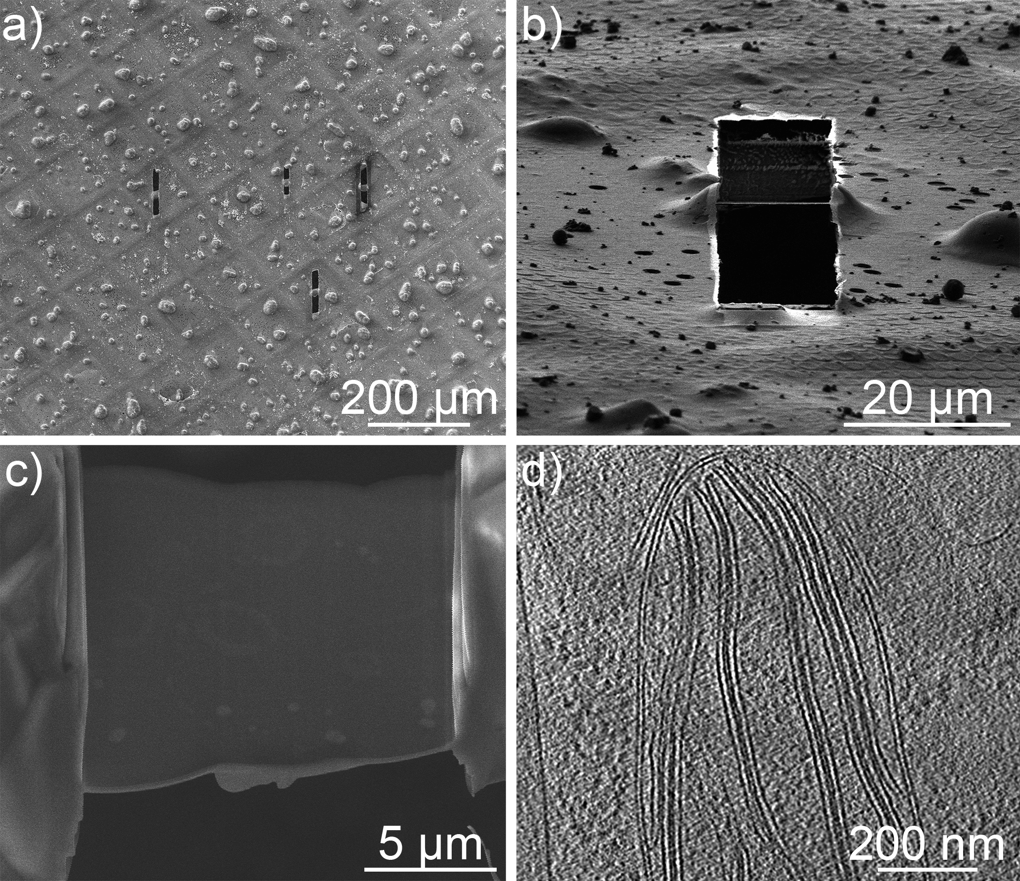
Fig. 1: Cryo-FIB preparation of Chlamydomonas reinhardtii cells. (a) An SEM image of a vitrified specimen on a TEM grid. (b) A FIB SE image of a lamella edge-on and (c) a top-down SEM image of a lamella. (d) A 2D slice from a tomographic reconstruction of a chloroplast within the intact cell, revealing the thylakoids and the chloroplast double membrane. |