IT-10-P-2061 Whole-Cell Imaging of the Budding Yeast Saccharomyces cerevisiae by High-Voltage Scanning Transmission Electron Tomography
High-voltage electron tomography provides three-dimensional (3D) information about cellular components in thicker sections beyond 1 μm, but image degradation caused by multiple inelastic scattering of transmitted electrons limits the attainable resolution. Scanning transmission electron microscopy (STEM) is believed to give enhanced contrast compared to conventional transmission electron microscopy (CTEM), and thicker samples up to around 1 μm can be analyzed with an intermediate-voltage electron microscope, because the depth of focus and the inelastic scattering are not the critical limitations.
We have applied STEM to 1 MV high-voltage electron tomography to extend the limitation of the specimen thickness, and seamlessly investigated the whole-cell structure of the budding yeast, Saccharomyces cerevisiae, size of which was ~3 μm width. The high-voltage STEM tomography, especially with a bright-field mode, demonstrated sufficient enhanced contrast and more-intense signals compared to regular TEM tomography (Fig. 1), permitting segmentation of major organelles in the entire cell (Fig. 2). The technique also showed less specimen shrinkage. The current spatial resolution is limited with the specimen preparation and the relatively large convergence angle of the scanning probe, but the present new technique has a potential to solve longstanding problems of image blurring in thick biological specimens beyond 1 μm, and to open a new research field in cell structural biology.
[1]K. Murata et al., to be submitted (2014).
This study was supported by the program of Joint Usage/Research Center for Develpment Medicine at IMEG, Kumamoto University. H-1250M at NIPS and JEM-1000K RS at Nagoya University were used for observation.
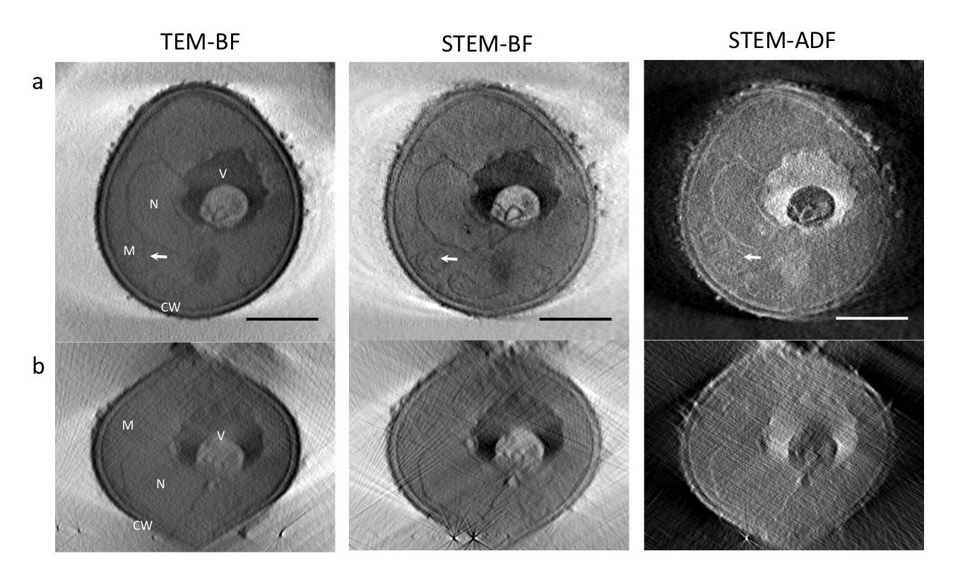
Fig. 1: Tomogram slices of an whole budding yeast cell at xy (a) and xz (b) planes were calculated from the tilt series collected by TEM-BF(Blight field), STEM-BF, and STEM-ADF(Annular dark field), respectively. Scale: 1 μm. |
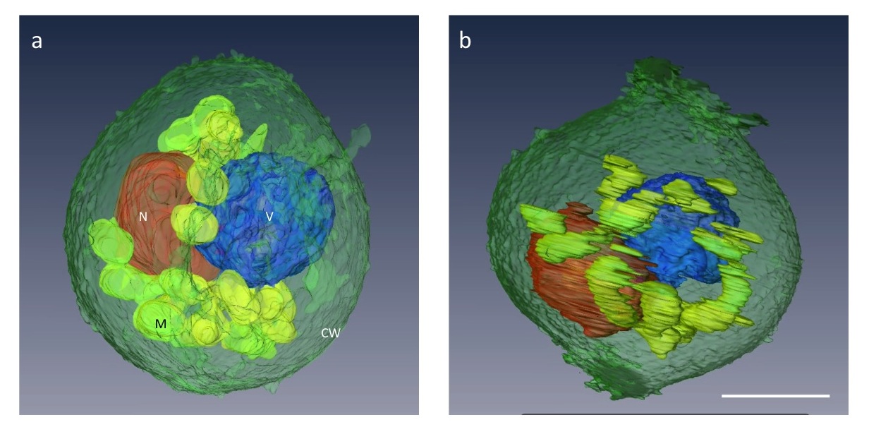
Fig. 2: The major organelles of a budding yeast cell were successfully segmented in a 3D tomogram calculated from the STEM-BF tilt series. Scale: 1 μm. |