IT-14-P-2022 A ferrule-top optomechanical probe to collect topographic and near field information about a sample at the nanometer scale
Atomic force microscopy (AFM) is an excellent technique for obtaining high-resolution images of the topography of a sample. The impact of AFMs in nanotechnology could be even more significant if the imaging capabilities were supported by an accurate mapping of the optical field in the close proximity of the surface. Scanning near field optical microscopy (SNOM), for example, has been shown to be able to combine AFM imaging with the possibility to collect optical information at the nanoscale. Yet, because of the complexity of its working principle, SNOM has been so far only used in specialized research laboratories and has been mostly limited to the analysis of surfaces in dry environments. To solve this limitation, in a previous work [1] some of us have proposed an all-optical device obtained by carving a tipped cantilever on top of an optical fiber. The opposite end of the fiber can be coupled to a readout system that was shown to be able to detect any tiny movement of the cantilever and to collect the SNOM signal coming from a prism illuminated under total internal reflection conditions. Here, we present another similar all-optical probe for AFM+SNOM imaging. The probe is based on ferrule-top technology [2-4], which relies on the possibility to fabricate a small cantilever on top of a ferruled fiber. This design keeps the overall advantages of the previous version (small dimensions, ease of use, easy integration in harsh environments) while significantly reducing the fabrication costs. Using this probe, we were able to obtain the SNOM profile and, simultaneously, the topographic image of a test grating (NT-MDT SNG01) kept in air and illuminated from below via an evanescent field. The AFM height resolution and the SNOM lateral resolution resulted to be comparable to conventional SNOM systems. Interestingly, measurements were repeated in water, with no major deterioration of the overall performance. This result paves the way for AFM+SNOM imaging on biological samples.
References:
1. B. Tiribilli, G. Margheri, P. Baschieri, C. Menozzi, D. C. Chavan, D. Iannuzzi, Journal of Microscopy 2011, 242, 10–14.
2. G. Gruca, K. Heeck, J. H. Rector, and D. Iannuzzi, Optics Letters 2013, 38, 1672.
3. G. Gruca, D. C. Chavan, J. H. Rector, K. Heeck, and D. Iannuzzi, Sensors and Actutators 2013, A190, 77.
4. D. C. Chavan, G. Gruca, S. de Man, M. Slaman, J. H. Rector, K. Heeck, D. Iannuzzi, Review of Scientific Instruments 2010, 81, 123702.
This work was funded by the European Research Council and by NanonextNL.
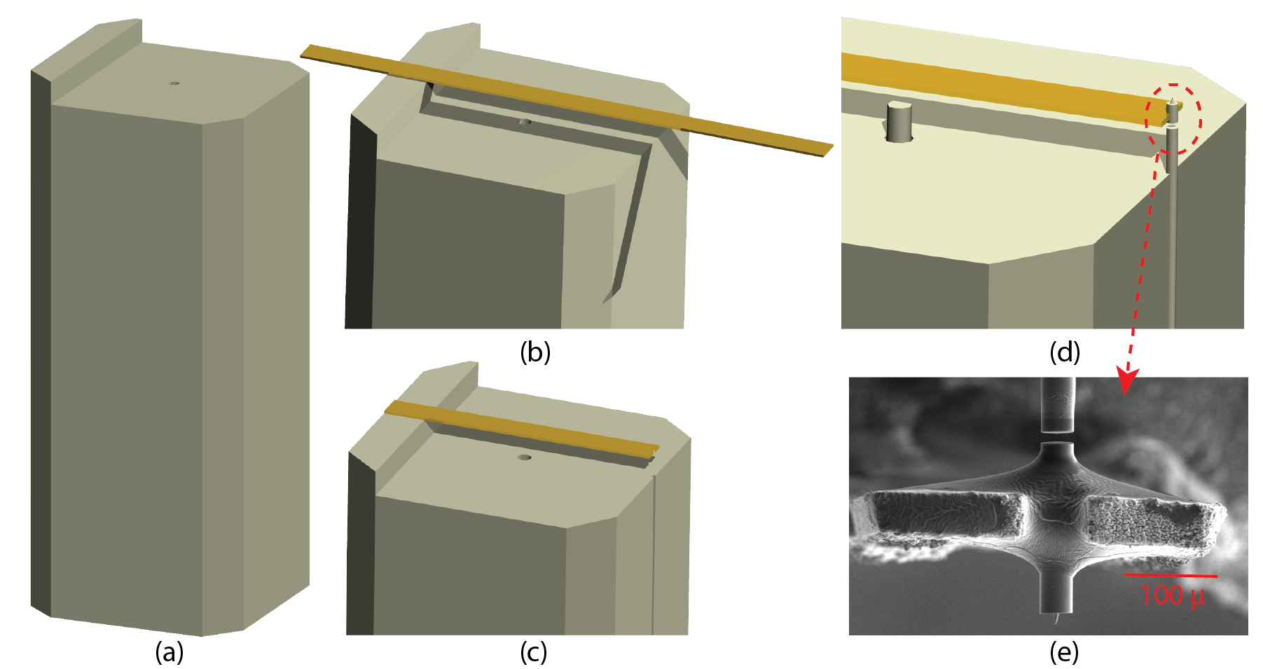
Fig. 1: Fabrication procedure of a ferrule-top probe. A glass ribbon is glued on top of a ferrule (a, b). The length of the cantilever is adjusted using a laser ablation machine (c). A tipped fiber is glued to the cantilever and the cantilever is released by focussed ion beam milling (FIB). Finally, a fiber is inserted into the borehole (d). |
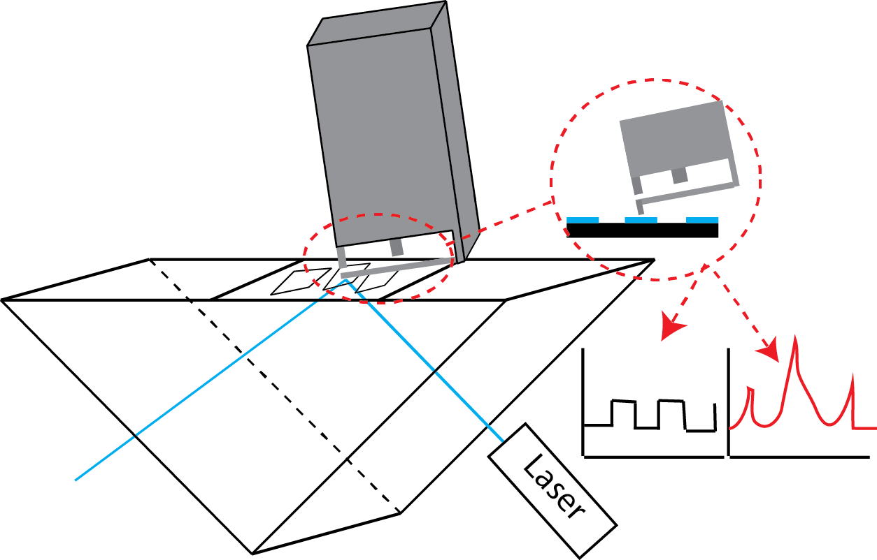
Fig. 2: A schematic view of the experimental setup. A blue (473 nm) laser beam impinges the top surface of a prism to create an evanescent field. The probe was scanned over the sample surface, in order to obtain a topographic and SNOM image simultaneously. |