IT-1-O-1982 Challenges in Phase Plate Development and Applications
While there have been attempts to implement phase plates in transmission electron microscopes (TEMs) over a long period of time, a publication by Danev and Nagayama [1] renewed interest that functional phase plates could be produced. In particular in life sciences, the development of thin film vitrification techniques has enabled the examination of unstained macromolecules and thin cells in the electron microscope, but also created the need for phase contrast. Conventionally, contrast at low spatial resolutions has been generated by using a strong defocus, but with the added consequence of introducing oscillations in the contrast transfer function. A phase plate allows one to work in-focus, with a large increase in the contrast at low spatial resolutions.
Many types of phase plates have been proposed, but the most widespread implementation has been the original thin-film Zernike phase plate. This type of phase plate has shown practical performance, especially in life science applications. The most widely tested film type is amorphous carbon, but these suffer from aging problems, making frequent exchanges of the phase plate necessary. Alternatives to conventional amorphous carbon have been investigated and silicon-based films show promise in terms of longevity.
In close collaboration with the Max Planck Institute of Biochemistry in Martinsried, FEI have developed a new type of phase plate with properties that make it very suitable for implementing it as a user friendly device in our TEMs. It produces high-contrast images, providing excellent contrast transfer in the low resolution range which is particularly relevant for cryo-electron tomography and may provide benefits for single particle analysis in the case of small and heterogeneous particles. No fringing effects around high-contrast features are observed and CTF oscillations can be avoided up to better than 10Å while maintaining contrast transfer at low spatial frequencies. Transmission losses by the phase plate are very modest. Moreover, the phase plate shows consistent performance for at least half a year of usage.
To facilitate routine phase plate usage we have added extra alignments and control panels to the microscope software. In particular, accurate adjustment of beam deflection pivot points is included to ensure a stable beam position at the plane of the phase plate. Also, software has been developed to easily navigate the phase plate in the back focal plane. We are developing detailed phase plate workflows for our applications software that will provide a seamless integration of the phase plate in the (automated) applications. In this talk a selection of results will be shown from cryo electron tomography.
[1] R. Danev, K. Nagayama, Ultramicroscopy 88, 243-252 (2001)
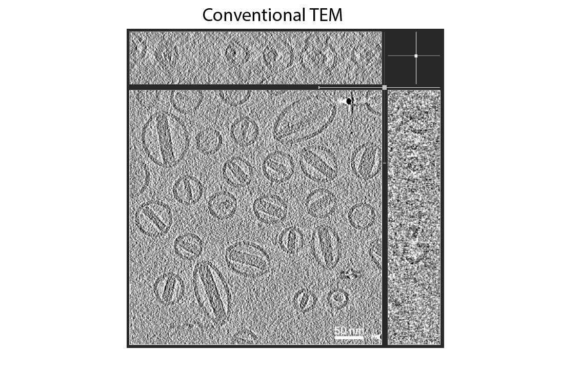
Fig. 1: Tecnai F20 results from cryo electron tomography on doxorubicin using conventional TEM at 4 μm defocus. Experimental conditions: total dose of 85 e-/Å2, tilt range +/-60º. |
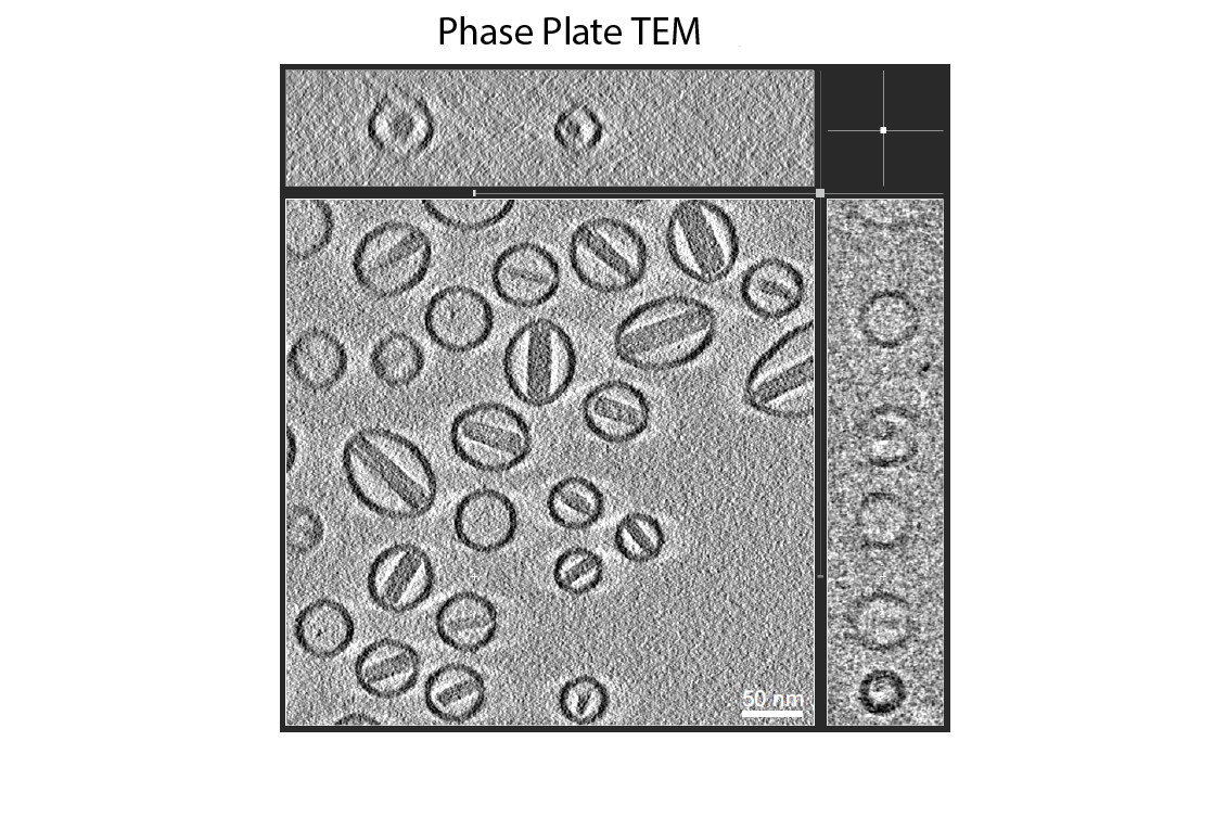
Fig. 2: Tecnai F20 results from cryo electron tomography on doxorubicin using phase plate TEM at 0.5 μm defocus. Experimental conditions: total dose of 85 e-/Å2, tilt range +/-60º. |