IT-6-P-1968 Investigation of hexagon shape nanoparticle growth mechanism using in-situ liquid Cell TEM
A number of efforts have been made on the synthesis of monodisperse nanoparticles with various morphologies to take advantage of their physical and chemical properties of nanoparticles attainable from the chemical composition and their dimensions. Growth of nanoparticle can be affected by many factors, such as temperature, surfactants, types of precursor, and its relative concentration. In order to understand the growth and the formation mechanism of nanoparticles, it was proven that the liquid cell TEM is one of the most powerful techniques because of its nanometer-level spatial resolution with transparency of the internal structure. Researchers were able to observe the growth procedures in real time using the liquid cell TEM, which made a great fore-step to observe nanoparticle growth using electron beam induced process.
Growth behaviors of spatially aligned, hexagon single crystals were investigated from the liquid cell with D.I water based solution. Formation sequence was observed from home-built liquid cell TEM stage in JEOL 2010F. No intentional heating was made during the crystallization. Streaming video was recorded from the Gatan ES500W camera. Figure1 shows the boundary regions with and without the nanoparticles formed by the electron beam induced growth. Inhomogeneous distribution of the particles might be come from exposure time difference for the particle formation. Figure2 shows the snapshots from the streaming video taken while shining electron beam onto the liquid layer. Nanoparticles were formed in spherical shape up to 123 second irradiation. However as irradiation time increased, nanoparticles were gradually changed to hexagonal shape. Orientation alignment and the growth rate were measured from the snapshots up to 260 second irradiation. Effect of precursor concentration and the electron current density on the formation and the growth of nanoparticles were examined.
This research was supported by the Nano-Material Technology Development Program(Green Nano Technology Development Program) through the National Research Foundation of Korea funded by the Ministry of Science, ICT & Future Planning (2011-0019984)
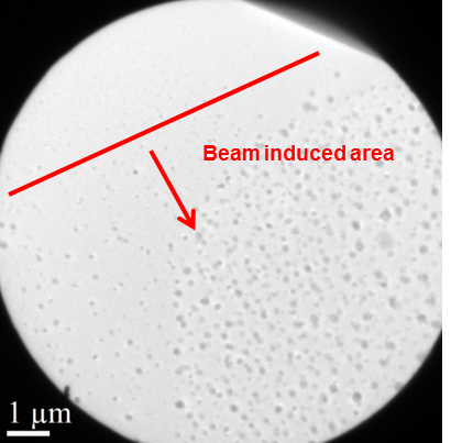
Fig. 1: Low magnification image of the field of view. The regions in which electron induced growth occurred or not. |
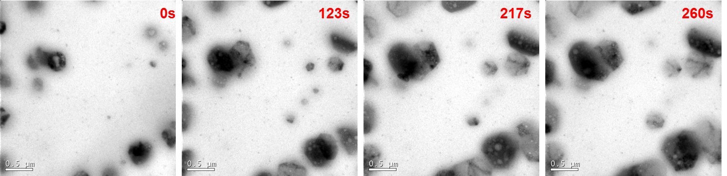
Fig. 2: Snapshots from the streaming video. As time increases hexagon shape nanoparticles growth were confirmed. |