IT-6-P-1924 Dynamic Observation of Gold Particles in Water by Environmental-cell TEM
An environmental transmission electron microscope (ETEM) is very powerful technique enabling to in-situ observation of specimens immersed in gases, electric/magnetic fields, and even in liquids by using a closed-type ETEM. In the closed system, diaphragms which are set at the top and the bottom of a space for the specimen to isolate from the vacuum are one of the most important components. Although silicon nitride (SiN) films are generally used for this purpose [1], charging effect on the diaphragms is inevitable because of the insulating SiN. In order to solve this problem, we have developed a carbon/SiN hybrid diaphragm which consists of amorphous carbon film as conductive base material and amorphous SiN thin layer coated on it [2]. In this study, the diaphragms were applied to in-situ TEM observation of motion of gold colloidal particles in water.
Figure 1 shows a photograph of our developed environmental liquid-cell. This consists of three parts; upper/lower grids and a spacer ring. Each of the grids had seven holes of ?0.1mm in diameter as windows for the electron beam, which were covered by the carbon/SiN hybrid membrane. The fluid specimen was sandwiched by these two membranes with controlling distance between them by 240 nm with the spacer deposited on the upper grid. The liquid-cell containing the fluid specimen was set in an environmental-cell specimen holder. In the experiment, the fluid specimen was gold colloidal particles having the size of 80±5.7nm in diameter dispersed in water in 1.1×10⁷/?l concentration, which were observed by using a conventional TEM H-8000 (200kV; Hitachi High Technologies).
Figures 2 show results of dynamic observations of motions of the gold particles in water. A gold particle in FIG. 2(a) migrated slowly at a speed of 10 ~ 30 nm/s, as shown in a quantitative analysis of variation of the moving speed in FIG. 2(b). This is not a Brownian motion because the gold particle drifted in a single direction. In contrast, a gold particle in FIG. 2(c) moved quickly just after starting the electron beam illumination at high speed of more than 1200 nm/s. Although a next short movement occurred after the first quick motion, the gold particle subsequently stopped, as shown in FIG. 2(d). These different types of the motion mean driving force of the movement of the gold particles is not unique. In the present cases, it is considered that an effect of water flow and coulomb force by charging due to the electron beam irradiation caused the motions of the particles in FIG. 2(a) and (c), respectively.
References
[1] U.Mirsaidov, C.D.Ohl, and P.Matsudaira, Soft Matter 8, 7108-7111(2012)
[2] T. Kawasaki et al., Proc. M&M2011. (2011) 465.
The authors acknowledge the financial support of this work by the Grant-in-Aid for Scientific Research (C) from the Japan Society for the Promotion of Science (#25390078).
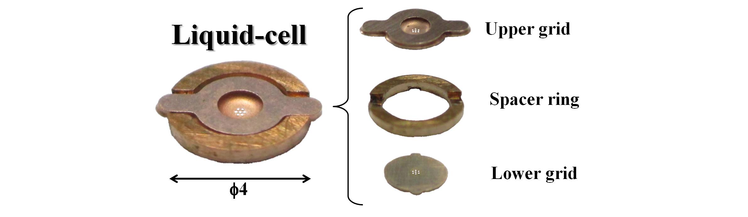
Fig. 1: Photograph of our developed environmental liquid-cell which consists of upper/lower grids and a spacer ring. |
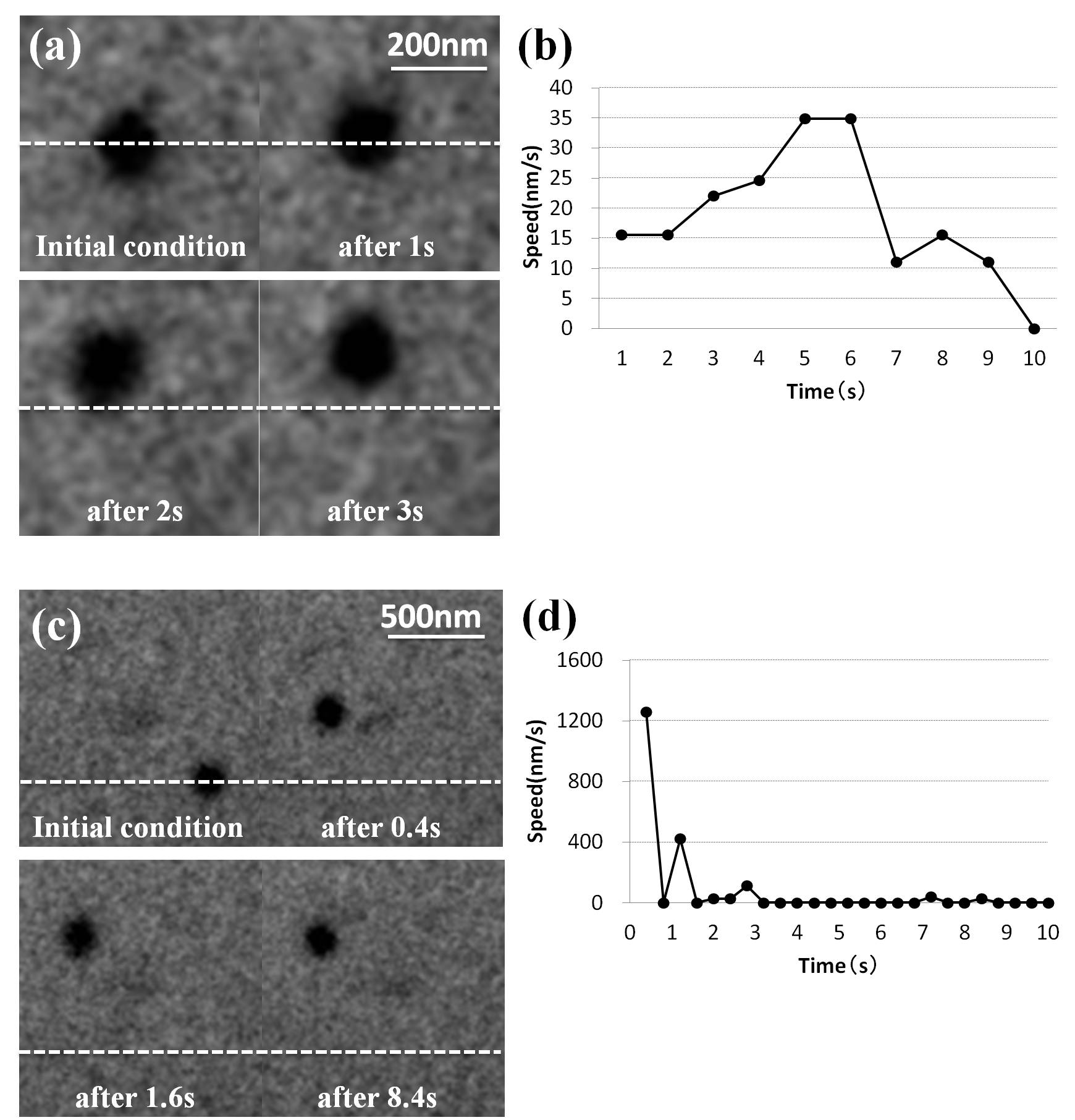
Fig. 2: TEM images of motion of gold particles ((a) slow and (c) quick movement) (b), (d) variation of speed of motions in (a) and (c), respectively. |