IT-13-P-1915 Advances in ex situ lift out
The focused ion beam (FIB) ex situ lift out (EXLO) technique for scanning/transmission electron microscopy (S/TEM) specimen preparation was historically the first lift out technique developed [1]. EXLO is well known for its ease, speed, and reproducibility, and is perfectly suited for manipulation of electron transparent specimens to carrier devices developed for in situ S/TEM testing as shown in figure 1a. Using EXLO for manipulation to a conventional carbon coated grid limits the specimen from being further FIB milled and inhibits certain S/TEM techniques. The development of a patent pending grid design and technique called EXpressLO™ allows EXLO and manipulation without needing a carbon film support [2-4]. The specimen is lifted out and manipulated directly to a slotted S/TEM grid surface such that the specimen may be directly analyzed and/or further FIB milled, broad beam ion milled or plasma cleaned. Using this new grid design, a specimen can also be manipulated easily into a backside orientation which avoids curtaining artifacts after further FIB milling [3]. The Xe+ ion plasma FIB (PFIB) is capable of producing electron transparent specimens for S/TEM [5]. The EXpressLO™ method can also be used for manipulating large PFIB prepared specimens as shown in figure 1b where a 50 micrometer long specimen is manipulated to a grid [6]. The 1 micrometer thick PFIB specimen manipulated to the EXpressLO™ grid can be further milled using conventional Ga+ ion FIB or a PFIB. EXLO is now flexible and continues to be fast and reproducible which saves labor and FIB instrumentation time, ultimately reducing the cost per specimen.
References:
[1] L.A. Giannuzzi, J.L. Drown, S.R. Brown, R.B. Irwin, F.A. Stevie, Mat. Res. Soc. Symp. Proc. Vol. 480, Workshop on Specimen Preparation for TEM of Materials IV, (1997), Materials Research Society, p. 19-27.
[2] L.A. Giannuzzi, Microscopy and Microanalysis 18, Supp 2, (2012) 632-633.
[3] Lucille A. Giannuzzi, Proceedings of ISTFA, ASM International. (2012), 388-390.
[4] L.A. Giannuzzi, Microscopy and Microanalysis 19, Supp 2, (2013) 906-907.
[5] L.A. Giannuzzi and N.S. Smith, in press, Microscopy and Microanalysis 20, Supp 2, (2014)
Qiang Xu from DENSsolutions provided samples and the in situ MEMS carrier device shown in figure 1a. Noel Smith from Oregon Physics provided the PFIB prepared specimen shown in figure 1b.
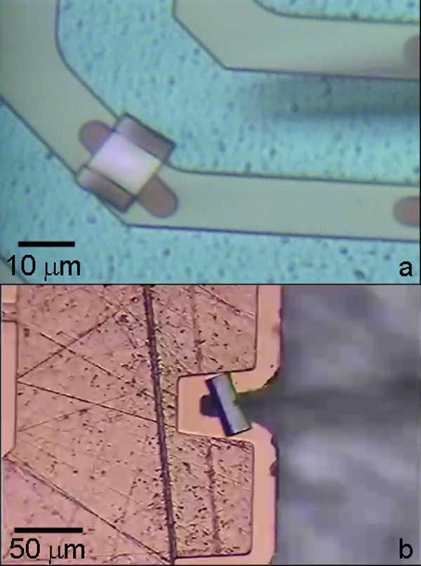
Fig. 1: (a) electron transparent specimen manipulated to a DENSsolution in situ carrier device via EXLO. (b) PFIB specimen manipulated via EXLO using the EXpressLO™ method and grid. |