IT-8-O-1901 Diffract-before-destroy with electrons?
Free-electron lasers provide x-ray pulses with short enough duration (< 100 fs) to record diffraction patterns from biological molecules (allowing their structure to be determined) before the molecules are destroyed by radiation damage [1]. Since fast electrons are elastically scattered more strongly than x-rays [2], it is reasonable to ask whether damage by radiolysis can be similarly outrun using electrons.
Electrons carry greater momentum than photons of the same energy, leading to additional knock-on damage, but in organic materials the knock-on damage is less severe than that caused by radiolysis [3]. More importantly, electrons carry electrostatic charge that can cause charging of insulating specimens (deflecting the beam or disrupting the specimen) and limit the incident-current density because of electrostatic repulsion between the electrons.
The Brookhaven ultrafast electron diffraction (UED) apparatus [4] produces 100fs pulses containing as many as 106 electrons at 2.8 MeV kinetic energy and this high energy helps to reduce Coulomb repulsion effects. To record ten diffracted electrons per pulse from a 10nm particle (e.g. macromolecule), the beam would need to be focused down to about 500 nm, giving a current density of almost 109 A/cm2. At these high current densities, space-charge effects are more important than statistical repulsion. Even so, it appears impossible to focus the beam to below a few micrometers diameter by means of a solenoid (Fig 1), due in part to the increased energy spread (approaching 0.3%) caused by Coulomb repulsion.
Lateral coherency of the beam is of concern for diffractive imaging, which nevertheless offers advantages over direct imaging since it avoids imaging lenses that degrade resolution because of aberrations and beam crossovers [5].
[1] JCH Spence, U Weierstall and HN Chapman, Rep. Prog. Phys. 75 (2012) 102601.
[2] R Henderson, Quarterly Rev. Biophys. 28 (1995) 171.
[3] RF Egerton, Microsc. Research & Technique 75 (2012) 1550.
[4] XJ Wang et al., J. Korean Phys. Soc. 48 (2006) 390.
[5] BW Reed et al., Microscopy and Microanalysis 15 (2009) 272.
Ray Egerton wishes to thank the Natural Sciences and Engineering research council of Canada for financial support. Renkai Li acknowledges DOE Grants No. DE-FG02-92ER40693 and No. DEFG02-07ER46272, and ONR Grant No. N000140711174. Work at BNL was supported by US DOE-BES under Contract no. DE-AC02-98CH10886
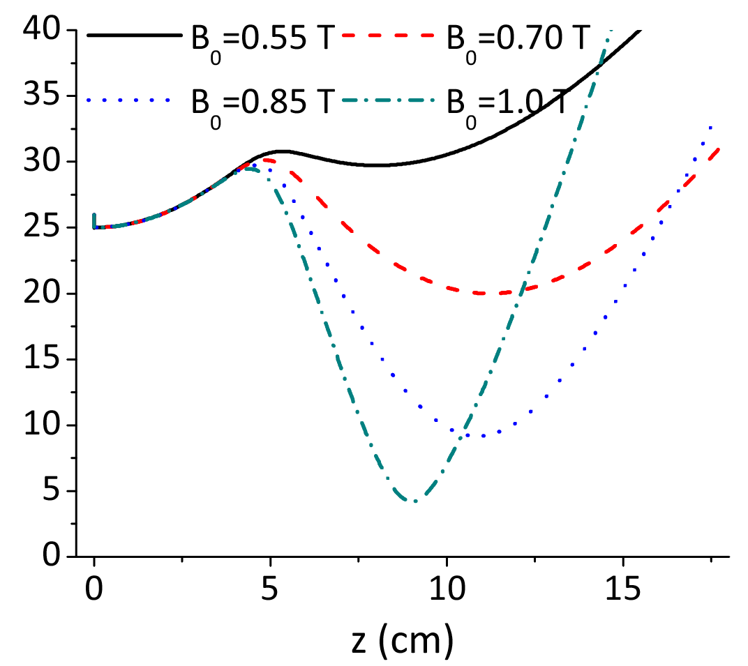
Fig. 1: Beam diameter versus distance along optic axis, calculated using the 3Dmesh method for a 0.16pC bunch of 2.5MeV electrons focused by a solenoid with four different values of maximum field strength B0. |
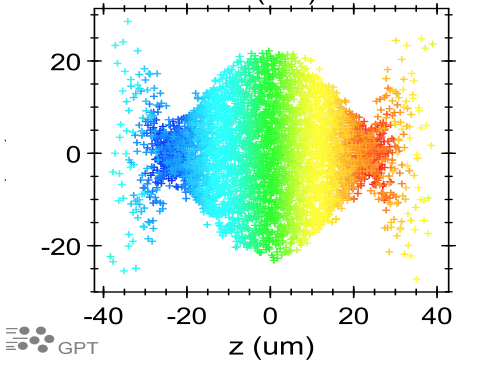
Fig. 2: Pulse profile, calculated using the 3Dmesh method and assuming an initial energy spread of 0.001%. Colors represent electron energy, red being the highest. |