IT-10-P-1875 Improving Depth of Focus in STEM Tomography using Focal Series
Scanning Transmission Electron Microscopy (STEM) is a point to point imaging method which uses a focused electron beam to build a projection image of the sample. One of the advantages of STEM is that the focused electron beam can pass through thicker samples (up to 1 µm-thick on a 200 kV FEG) providing higher signal-to-noise ratio than standard TEM (wide spread beam). This makes STEM more suitable for electron tomography of thick samples. Nevertheless, the smaller is the size of the probe, the more reduced is the depth of focus (DOF). A way of solving this difficulty is to take advantage of raster scanning to get focused images line by line by dynamic focus [1]. However, in our experience, the recovery of full-focused images by dynamic focus is not helpful when the DOF cannot encompass a very thick sample (>0.5 µm). To circumvent this limitation we have developed an acquisition scheme and an image processing method in which we reconstruct full-focused images from STEM images recorded at different defocus.
This process of DOF correction can be used for both 2D studies and tomographic experiments. Briefly, it consists in two steps. For a 500 nm thick sample, i) five images are acquired at different focus values ranging from -300 nm up to 300 nm defocus (using 150 nm steps); ii) the images are processed using an ImageJ macro based on Turboreg [2] and Extended Depth of Field [3]. This macro consists of two steps: i) the images acquired at different focus are aligned; ii) regions of these registered images which are at focus are combined to compute a single image.
The performance, in terms of resolution, contrast and SNR, of the afore mentioned approach has been evaluated in silico by comparing 3D reconstructions computed from the projections of a phantom volume leading to two different datasets. In the first dataset the whole images are at focus whereas in the second one, parts of the images have been modified to mimic the focus changes observed in experimental data. The method was applied to compute the 3D reconstruction of a resin-embedded T. brucei 500 nm-thick section using five focal series. Sections of the reconstructed volumes without (1 focal series), with intermediate (3 focal series) and full (5 focal series) DOF correction on high-tilt images (±40°) are displayed in figure 1 showing the improvement in the observed details.
References:
1. Feng et al. “Automated electron tomography with scanning transmission electron microscopy”. J. Microsc., 2007, 228:406-12.
2. Thévenaz et al. "A Pyramid Approach to Subpixel Registration Based on Intensity". IEEE Tr. Im. Pro., 1998, 7: 27-41.
3. Forster et al. « Complex Wavelets for Extended Depth-of-Field: A New Method for the Fusion of Multichannel Microscopy Images”. Mic. Res. Tech., 2004, 65:33-42.
This work has been funded by the ANR grant 11-BSV8-0016.
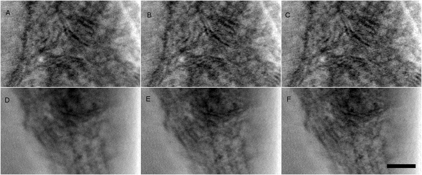
Fig. 1: Comparison of 3D information recovered from depth of focus correction of high-tilt images. a, b, c) 1 nm-thick XY planes from reconstructions computed with only high-tilt images (±40°) without, with intermediate and full depth of focus correction respectively. d, e, f) 1 nm-thick YZ planes from the same reconstructions. Scale bar, 100 nm. |