IT-13-P-1838 FOCUSED ION BEAM LITHOGRAPHY OF SUBWAVELENGTH PHOTONIC 3D-CHIRAL STRUCTURES
The focused ion beam (FIB) milling is a powerful tool for fabricating nanoscale photonic structures [1]. As the next step after successful fabrication of lamellar optical gratings with subwavelength periods [2], we employ the FIB technique to produce truly 3D patterned nano-scale structures. The report describes fabrication and analysis of periodic subwavelength arrays of 3D-chiral holes in a freely suspended silver film.
In order to generate digital templates for FIB lithography (‘stream files’) a special numerical routine has been developed. The templates contain the ion beam waypoints’ coordinates and their ‘dwell time’. Accordingly, all the desired characteristics such as the form, dimensions and the etching depth of a single element can be set. Employing this method allowed us to fabricate periodic arrays of 3D-chiral holes in the freely suspended 200 nm thick silver film with a total processed area of 30x30 μm2 and one element size of 300 nm. Fig. 1a features a 3D lithography model of a single chiral element. The X, Y, and Z coordinates correspond to those of waypoints in the template and the dwell time, respectively. Fig. 1b shows a micrograph of the fabricated structure tilted by 52°, and Fig. 1c shows a normal view of the structure. The fabricated structures have proven to exhibit significant optical activity and circular dichroism.
[1] C. Enkrich, F. Perez-Williard, D. Gerthsen, J. Zhou, T. Koschny, C.M. Soukoulis, M. Wegener, S. Linden, Focused-ion-beam nanofabrication of near-infrared magnetic metamaterials, Adv. Mater. 17 (2005) 2547.
[2] M.V. Gorkunov, V.V. Artemov, S.G. Yudin, S.P. Palto, Tarnishing of silver subwavelength slit gratings and its effect on extraordinary optical transmission, Phot. Nanostr. Fund. Appl. (2013) http://dx.doi.org/10.1016/j.photonics.2013.10.001.
This research was financially supported by the RFBR No. 13- 02-12151 ofi_m and the RAS Presidium program 24. We are grateful to A. L. Vasiliev for the access to the FEI Helios microscope.
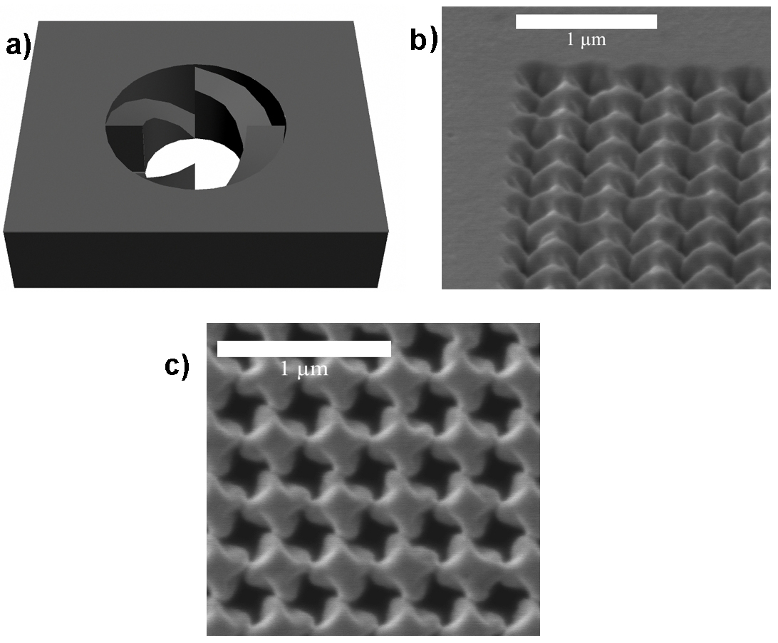
Fig. 1: 3D model of the chiral structure unit cell as implemented into the FIB milling digital template (a), SEM micrograph of the fabricated structure tiled by 52° (b), normal view of the fabricated structure (c). |